


New microscope to see intracellular activity in 3D and in real time
Today a new optical microscope has been presented capable of visualizing the biological processes that occur at high speed and on a small scale. From the development of an embryo to the movements of individual proteins, the microscope collects in real time and 3D what is happening. Researchers at the Howard Hughes Medical Institute have presented the new microscopy technique in the journal Science. In fact, one of the 2014 Nobel Prizes in Chemistry leads the research team that has developed the technique: Eric Betzig.
Microscopy techniques for visualizing biological processes in vivo have two limitations. On the one hand, when zooming for very small scale high-resolution images, the sample is damaged by the light emitted for observation. On the other hand, they need more time to take pictures, limiting the ability of microscopes to follow rapid processes.
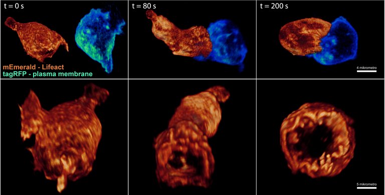
The new technique presented by the Betzigen group considerably reduces the damage to the sample and increases the list of biological processes that can be studied under an optical microscope. The capacity of the new microscope has been tested with 20 biological systems, obtaining spectacular images and videos. The following video shows, for example, the muscle contractions of a worm embryo C. elegans:
Movie S6 High Resolution from HHMI NEWS on Vimeo.
The new technique has been created from light sheet microscopy. This technique involves using lasers to form light layers and illuminate the sample by sections. The thinner the light layer, the higher the resolution of the microscope. This microscopy technique has been used to follow in vivo the development of embryos, but the layers were too thick to follow smaller scale processes. Giving the light layers a reticular structure, the Betzig team has invented the way to create layers of ultraviolet light. This has invented the way to reduce sample damage and increase speed resolution. The name of the new technique is Lattice Light Sheet Microscopy, and researchers who want to test it should request it at the following address: http://www.janemia.org/aic
Buletina
Bidali zure helbide elektronikoa eta jaso asteroko buletina zure sarrera-ontzian


