


Two brain maps, the first fruits of the BRAIN project
The BRAIN project was launched in the spring of last year with great intentions: to create a map of brain functioning in healthy and sick people. They have three main objectives: to know how the brain processes information, to understand the function of cells at a genetic level, to identify and classify all types of cells that make up the brain, as well as their relationships. The project, led by the Allen Institute for Brain Science, has numerous international researchers, and proof of this are the published articles.
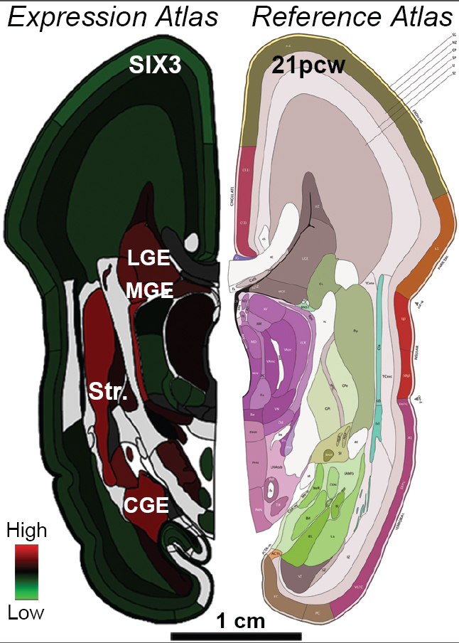
It should be noted that all the data obtained are public. Thus, researchers have advanced that the Atlas on the brain of the human embryo presented in the first article will be of great use to many scientists. Thomas R confirms this idea. Director of the Mental Health Institute of the United States: "This atlas is already changing the evolution of the human brain and how to investigate diseases related to neuronal development, such as autism and schizophrenia," he said.
In fact, in adults there are genes connected to each other in the brain of the embryo. Therefore, understanding the functioning of these genes (when they are expressed, when they are not, what relationship they have between them) is key to knowing the future development of the child's brain and the young man after birth. The map presented includes this operation.
In the investigation of autism, for example, it begins to bear fruit. Besides being very useful in this aspect, they have emphasized that it helps us to understand in what is distinguished our species of other animals. According to Ad Lein, researcher at the Allen Institute, "we know that there are areas of the human genome that show surprising differences with other animal species. Since it can give clues about the function of each of the genes expressed in the brain, we can use our map to understand what makes our brain special."
Map of neuronal mouse connections
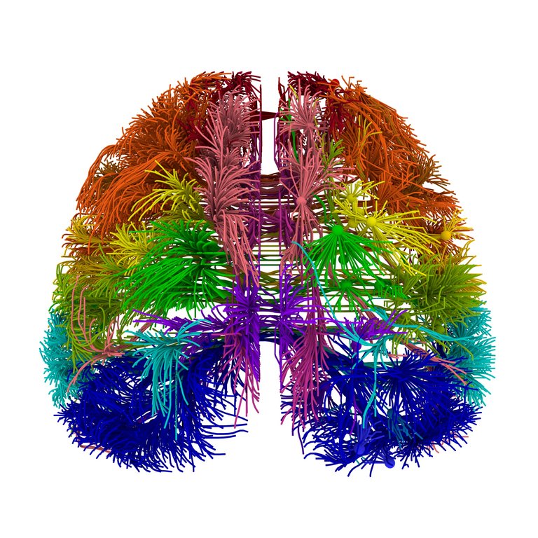
The second article helps to investigate how the brain processes information. Specifically, they have completed the map of how mouse neurons are connected: Mouse Brain Connectivity Atlas. This is a very big step, since the only complete map so far was that of nematode C. elegans (C. elegans has 302 neurons, mouse 75 million and us 100,000 million).
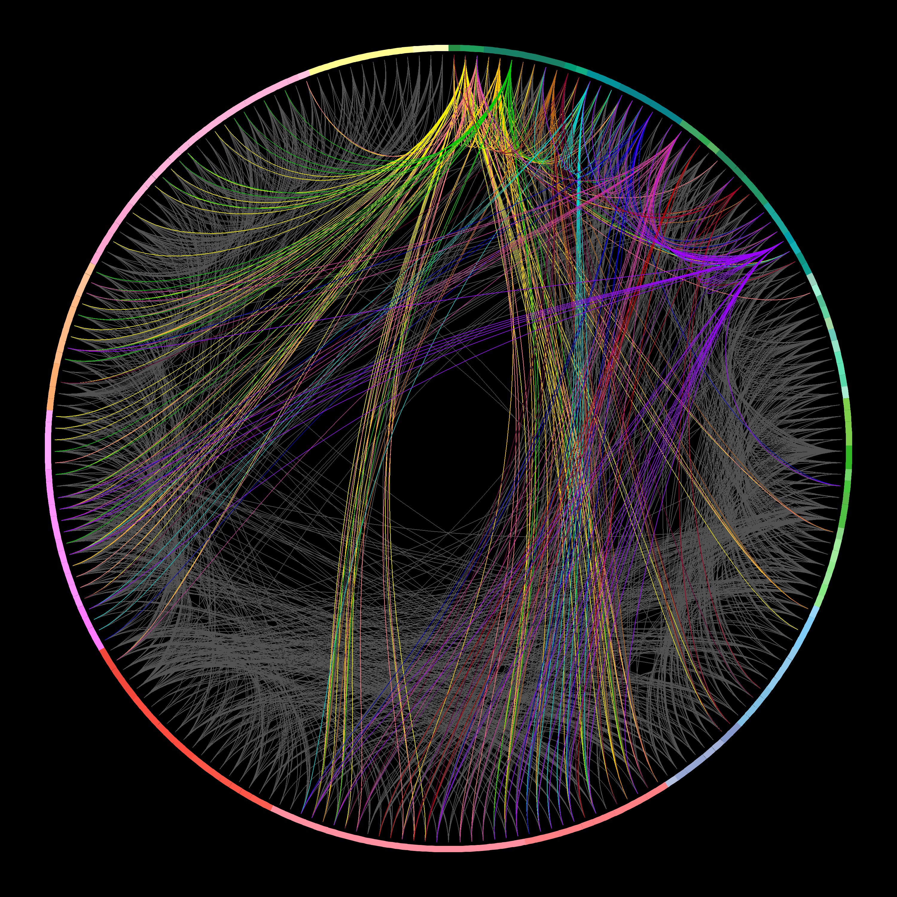
As the researchers have explained, when completing the maps of the nematode and the mouse, the difference has not only been in the number of neurons, but has methodologically taken one more step. Specifically, genetically modified viruses have been used for the individual monitoring of neurons. As for the location in three dimensions, a standardized program has been developed that has allowed to process so quickly the total amount of data generated (1.8 petabytes).
Buletina
Bidali zure helbide elektronikoa eta jaso asteroko buletina zure sarrera-ontzian


