


Radiological protection... essential
Ionizing radiation
Electromagnetic radiation is classified into two large groups: non-ionizing radiation and ionizing radiation. The limit between the two is a certain energy level. Non-ionizing radiation has a lower energy level and is visible radiation, infrared rays, microwaves and radio waves. Ionizing radiation, on the other hand, has a higher energy level and can be both atomic particles (alpha and beta particle radiation) and electromagnetic (ultraviolet rays, x-rays and gamma rays).
Today it is known that non-ionizing radiation warms the body's tissues, but it has not been shown to cause other potentially harmful health effects. Ionizing radiation can penetrate matter and cause atomic alterations.
Since atoms are basic components of tissues, changes in atoms can cause various tissue damage. It is true that the tissues have the capacity of self-protection (example of this is the melanin that we have to protect themselves from the sun's rays), but that protection is only of a certain magnitude; from there occur somatic damage (when the radiation receptor suffers the disease) or genetic damage (when mutations occur in the genes and affect the following).
Applications

The uses of ionizing radiation are very diverse: carrying out X-rays, cancer therapy, measuring thickness, density or humidity of materials, fire detection, sterilization of medical equipment, elimination of pests, irradiation of food, soil fertilization, study of layers in geological prospecting or archaeological dating, among others.
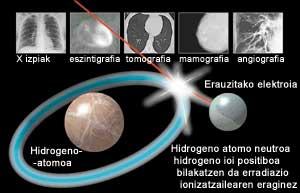
X-rays or gamma rays are known among medical uses, but many nuclear techniques are used in hospitals. Chemical compounds mixed with low activity radioactive elements, for example, are usually used for diagnostic purposes: once the ‘marker isotope’ is injected into the body, thanks to the radiation it emits, doctors inspect the movement of the isotope in the organ they want to investigate, in order to know if its functioning is correct.
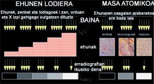
Depending on the thickness, density or composition of body tissues, the absorption of ionizing radiation as the body is traversed is different. For example, calcium in bones has an atomic mass greater than hydrogen in tissue water. This difference means that when the X-rays cross our body, they are eliminated more strongly in the bones and, therefore, the ‘shadow’ of the bones is clearer in the x-ray image. Even when the organ that passes through the radiation is thicker, its ‘shadow’ is better distinguished. Therefore, in X-rays the ‘shadows’ give us information about the internal structure, the ‘shadows’ that, once the radiation has crossed our body, form bones or other organs.
Biological damage to radiation
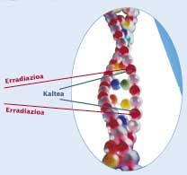
Ionizing radiations when they reach the cells of living beings produce harmful genetic or somatic effects. In the case of somatic damage, the disease is suffered by the radiation receptor itself; in the case of genetic damage, mutations occur in genes and diseases occur in descendants. The magnitude of the effect depends on the type of radiated cell, the absorbed dose, the exposure time, the radiation energy and the penetration capacity.
Alpha radiation is little penetrating. Beta radiation, like alpha, ionizes the medium it passes through, but is much more penetrating than it is. X-rays, on the other hand, are very penetrating and can cause serious damage to the body (tissue destruction, skin burns, DNA damage, etc. ). Finally, gamma rays are the most penetrating and therefore the most dangerous radiations.
The biological effects produced by radiation are measured by equivalent dose and their unit is sievert (SI). If the doses received are high, the damage can be immediate. In low doses, damage may appear longer term.

There are two types of effects: stochastic and non-stochastic. In some cases, the damage that radiation can cause increases with the dose, is the case of stochastic effects. Suppose we play in lottery (‘prize’: cancer, hereditary disease…); it is clear that the probability of getting the winning lottery ticket (damage) increases as we buy the tickets (dose) and that we have the chance to win from the first ticket we buy. On the contrary, if there is a threshold dose, that is, if there is no damage until reaching that threshold dose, and from that threshold dose, if as the dose increases, the damage can also increase, we talk about non-stochastic effects.
Following the example of the lottery, in this case no lottery tickets are bought from the beginning. Limiting non-stochastic effects is very simple, as that threshold dose will be the limit. The limitation of stochastic effects is based on statistics. There is a statistically measured risk for all jobs. This data is known. To limit the risk of radiation-related jobs the average of the rest has been done. have been based on statistical studies conducted in populations of Hiroshima and Nagasaki to relate this risk to a certain level of radiation.

This has limited the maximum level of radiation a person can receive. However, normally these levels are much lower (both limits and levels actually perceived). However, there is no evidence that low dose damage can occur.
New ionizing radiation protection regulations
The radiological protection regulations are based on three principles: justification, optimization and limitation. The principle of justification states that the benefit obtained from ionizing radiation is the only cause of exposure to them. The second, the optimization principle, states that doses should be as low as possible, also called the ALARA principle (As Low As Reasonably Achievable). And, according to the third, doses should be limited and these limits materialize in the regulations.
For regulatory development in this area, States follow the international recommendations published by the ICRP (International Commission on Radiological Protection). ICRP is an international working group that brings together specialists in radiological protection, publishes recommendations and states adapt these recommendations to their regulations.

The new legislation is based on the recommendations published by the ICRP in 1991. The new regulation has allowed a greater limitation of doses, the elaboration of more realistic metabolic models for the calculation of internal doses and the introduction of natural radioactivity. But there is room for improvement, and without forgetting the collective dose limit, experts have pointed out the need to further differentiate individual doses. As for the general public, it has been recommended not only simplification but also differentiation, and that environmental protection should be integrated into the regulations. It seems that the ICRP will publish new recommendations for 2005.
What are ionizing radiation?
Electromagnetic radiation is classified into two large groups: non-ionizing radiation and ionizing radiation. The limit between the two is a certain energy level. Non-ionizing radiation is visible radiation, infrared rays, microwaves, and radio waves. Ionizing radiation, on the other hand, has the ability to pass through matter and ionize neutral atoms (losing the electrical balance), being both atomic particles (radiations of alpha and beta particles) and electromagnetic (ultraviolet rays, X-rays and gamma rays).
Alpha and beta radiation is transmitted by the disintegration of radioactive substances and the release of beta radiation into nuclear fission. Alpha radiation consists of helium nuclei with two neutrons and two protons, while beta radiation consists of electrons. In medicine, beta radiation is used in cancer radiation therapy treatments.
Another important particle radiation is cosmic rays. It is a radiation formed by particles of great energy that expand in space, mainly protons and helium nuclei. When the Earth's atmosphere falls, it becomes a radiation formed by elemental particles and gamma rays. The main causes of cosmic rays coming to Earth are the Sun and the center of the galaxy.
X-rays are emitted when an electron from the inner orbitals of the atom leaves the atom (emits the electrons from the outer layers that will fill the inner gap). They are formed in vacuum tubes (x-ray tubes) and are mainly used for performing X-rays (in medicine, industry, still).
Gamma rays form an electromagnetic radiation of lower wavelength and therefore of greater energy. Like alpha and beta radiation, they are emitted by disintegration of radioactive materials and fission of fissionable materials. Its main use is medicine, scan and radiation therapy. High-frequency gamma radiation is a small part of the cosmic rays that reach Earth through supernovae or other galaxies.
Influence of ionizing radiation on life
Ionizing radiation causes changes in living tissue that can damage or even kill cells. If the exposure is sufficiently high and therefore affects a large number of cells, the effect can range from the combustion of the skin to produce more harmful and/or harmful effects. Radiation exposure also increases the risk of developing cancer.
And around us, what? Currently, in the Autonomous Community of the Basque Country there are 110 facilities generating ionizing radiation. These include medical facilities (radiation therapy, nuclear medicine and cobaltotherapy), industry (especially in steel and metallurgical companies) and university research and education laboratories. According to the agreement signed in April between the Basque Government and the CSN (Nuclear Safety Council), the Government of Vitoria-Gasteiz is responsible for the inspection, evaluation, control and transport of materials and the radiological surveillance of the environment. For more information on this topic you can consult the website www.euskadi.net/vigilanciaradio. |
Geoffrey Webb: “In the regulations it will be necessary to differentiate more the limits of the doses and take into account the environment”
What are the main sources of radiation today and where are they generated?
The first thing to be made clear is that people do not realize that we live in a radioactive world, and most of it is cosmic radiation and that coming from the rocks under our feet. For example, the uranium we hear so much comes from the underground.
We naturally live in a place full of radiations. Man, with his radiological works, adds an extra to these natural doses. We, from our association, try to keep these extra doses under control.
Therefore, we can say that there are two types of radiations: natural and human radiation.
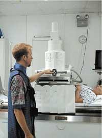
So it is. The most important source of these extra radiations produced by humans are hospital X-rays, a worldwide source of radiation. It is the main source of radiation in developed countries, much higher than that resulting from nuclear power plants.
How to protect yourself from these radiations? What measures should be taken?

First you have to measure radiation and then establish laws.
In the case of medicine, doses are set for each type of treatment and diagnosis. That is, the radiation applied in each treatment does not exceed a quantity.
It is not possible to establish a fixed dose, since the dose necessary according to the characteristics of each person can be very variable. We try to establish quantities that serve as reference for each treatment.
The opposite happens with nuclear force. We, in this case, set limits: a person working at a nuclear power plant cannot exceed the radiation limit we set annually.
And finally, there are people outside the nuclear power plants, which we call public. For them we also set limits on the amounts of radiation they can receive annually. Needless to say, the limits set for the public are much lower compared to the people who work in the power plants.
When setting these limits we always consider the so-called ALARA principle. ALAR means: ‘As Low As Reasonably Achiveable’. That is, if a person, for his work or for health problems, needs to undergo radiation, he will receive the minimum amount of radiation needed to obtain a suitable result.
You are now president of IRPA. What is your main function?
In short, analyze the biological damage of radiation.
There is some danger in everything that human beings do, nothing is inexorable, so we try to identify all possible risks. Then we try to set radiation emission limits compatible with the work. Once this is done, they turn to the governments of the countries and are given those councils to legislate for compliance.
We could summarize, therefore, that our association works directly with professionals from countries to develop the regulatory regulations of each country.
Do you think countries meet those limitations that you have established?
In Europe and North America yes. Countries with very strict laws and regulations. But in developing countries, Africa, South America and some places in Asia is much more difficult.
Something special happens with the states of the Soviet Union: they have their laws, but they have become obsolete. Until recently I have worked with scientists from Estonia, Latvia and Lithuania to create new legislation.
This year the new regulations for protection against ionizing radiation have been implemented. Where are the following recommendations going? What needs to be improved?
On the one hand, when delimiting doses, it is necessary to go to differentiation, that is, to see and analyze each case individually. For example, it is not the same, in the case of X-ray treatment, to take an x-ray of a thick person or a child. The former needs a greater number of radiations to obtain the same result. Doses should be adapted in each case.
On the other hand, it is necessary to integrate the radiological protection of the environment in the regulations, as is being studied.
Buletina
Bidali zure helbide elektronikoa eta jaso asteroko buletina zure sarrera-ontzian


