


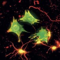
Astrocytes in the head
One way to investigate brain function is to study electrical activity. Neurologists have developed this type of research a lot. The brain is continuously passing electrical signals through neurons. It is the engine of a car standing but running; the brain is working. But when the body does something, sees, hears, moves, ole, feels, or launches another type of activity, the flow of cerebral electric signals increases in certain areas. Neurologists seek the correlation between the activities of the body and the parts of the brain that are activated. They are getting amazing results.
But this is not the only way to understand how the brain works. Another way is to follow the electrical signals at the cellular and molecular level and is gaining strength. This is also not a new line of research; for some time now we are studying what happens with the electrical signal to be able to jump from one neuron to another, where and how electrical signals are generated, etc. But, despite its antiquity, this path has not been exhausted at all. Precisely, state-of-the-art techniques have opened new doors. New questions. And over time, research into non-neural components has become essential, including astrocytes.
Energy demand
The question that has led researchers to deal with astrocytes has to do with energy. Where do neurons extract the energy needed to transport the electrical signal? Some molecules are known to transport the signal in exchange. But the synthesis and mobilization of these molecules consumes energy. Where does that energy come from?
As in many cellular processes, this energy comes from the oxidation of glucose, ultimately from the molecules that the blood brings. When the body does something, neurons consume more energy than when it is stopped, meaning it needs a lot more blood, more glucose and more oxygen. The numbers are spectacular, the brain has only 2% of the body mass, but it consumes 15% of the blood, requires 25% of glucose consumption throughout the body and approximately 20% of oxygen consumption.
Depending on the needs, neurons take glucose and oxygen from blood vessels, produce oxidation, generate energy and move neurotransmitters.
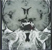
For all this to be effective, the brain needs a solid infrastructure. Researchers say that the brain is a very vascular organ, that is, with a very high density of small blood vessels compared to other organs. It has a compact network of capillaries that allows glucose and oxygen to reach all neurons through the blood. In short, the brain works thanks to two components: neurons and blood vessels.
However, there is a problem; if we look closely, we see that blood vessels and neurons are not together. Blood does not release glucose or oxygen directly into neurons. Among them is a third component, astrocytes. In view of this, the conclusion is correct: the work of astrocytes is to communicate blood vessels and neurons and carry out transport work.
More than support
The function of astrocytes is not evident. Even the development of existing fine techniques has not been possible to analyze. In addition, there was no need because neurologists considered that only neurons have to do with brain activity. It is believed that the goal of all types of cells was to maintain the structure of the neural network. In fact, pathologist Rudolf Virchov called glia other non-neuronal cells, derived from the English word glue. Actually, glia is a mixture of several cells, but the most abundant are astrocytes. In addition, they are as abundant as neurons.
Today, it is a large cast of well-known astrocytic functions. In addition to the communication between blood vessels and neurons, and in addition to serving as support, they perform many other functions, most related to the molecular control of the area. In short, they do the cleaning, expel dangerous molecules, etc.
These tasks also consume energy, it is not a great consumption, especially compared to the one that consumes neuronal activity. However, to understand the energy balance of the brain it is necessary to investigate the dynamics of astrocytes, which work with neurons.
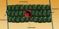
In fact, astrocytes are physically attached to the other two components. Thus, the neuronal environment detects the activity of the synapses and coordinates with that of the blood vessels to respond to this activity.
Glutamate
A high percentage of synapse activity is related to a single neurotransmitter: glutamate. In the cortex, that is, in the part of the brain that performs most cognitive functions, glutamate represents more than 80% of neurotransmitters. It is the most common neurotransmitter. That's why your metabolism is used to study brain activity.
The synapse emits glutamate and, after transmission of the signal, astrocytes recycle it. The process is extremely fast. For two reasons. On the one hand, the accumulation of glutamate in the brain is very dangerous, since excessive stimulation of neurons can cause a heart attack. To transmit the signal just a little glutamate and the rest will be removed quickly. Moreover, excess glutamate produces a background sound in signal transmission. It is evident, therefore, that the elimination of excess glutamate must be rapid, work performed by astrocytes through the recycling of glutamate, transformed into another molecule by means of chemical reactions, to convert it back into glutamate in neurons.
This work consumes energy. It is not much, but astrocytes have to recover that energy. A small proportion of the glucose they take from the blood is directed to it. Precisely, the techniques of representation of the activity of the brain perceive this energy consumption and, ultimately, detect the activity of the brain, since the work of neurons and the energy consumption of astrocytes occur jointly.
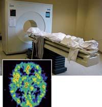
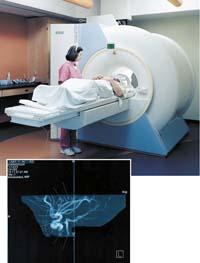
Networks
Internal brain scanning techniques are techniques for detecting metabolic changes. For this reason, the user must know in detail the metabolic process that is reading the technique in each experiment.
The glutamate removal process is an example. And a very fruitful example, since the research of this metabolic pathway has brought surprises.
Neurologists at the University of Lausanne have discovered that networks are formed not only by neurons, but also by astrozios. In fact, communication between neurons and blood vessels occurs as a wave in astrocytes. It is a metabolic wave, that is, glucose, oxygen and other substrates extend to all astrocytes that surround the blood vessel, and from them to those around it, etc. They extend in all directions.
Therefore, communication does not occur in a straight line, but extends to a whole space; the process that is initially activated in a few synapses also extends to other beliefs of the environment. In this way, communication between two connected neurons can launch other nearby neurons even if they are not directly related.
These mechanisms make it more difficult than expected to understand the functioning of neural networks. In addition, many other functions of astrocytes may be unresolved.
Much work is lacking in astrocyte research. For example, some MIT neurologists have proposed that the culprit for some diseases is not necessarily a failure in the functioning of neurons, but that failures in the astrocyte network will also have to do with those diseases. According to them, these diseases could include autism and schizophrenia.
Otherwise, in some experiments astrocytes do not always supply neurons with all the energy they need. In these cases neural losses occur. It is unclear why this happens, and brain scanning techniques may need to be improved to answer these questions. Neurons are very interesting, but if you look well you see other interesting cells in the brain. Yes, you have to be able to look good.
Buletina
Bidali zure helbide elektronikoa eta jaso asteroko buletina zure sarrera-ontzian


