
 Bilatzailea


Photo of the week: herpes on the skin
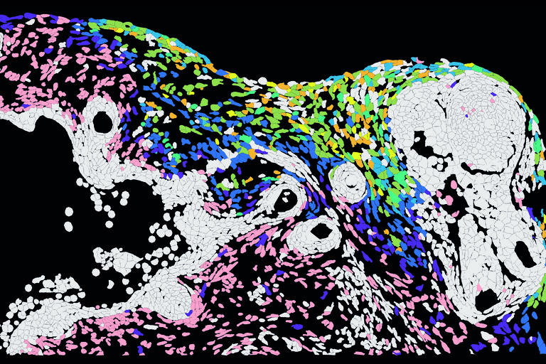
Researchers at the Max Delbrück Center have studied the spread of herpes virus type 1 infection using skin organoids.
Each colored cell in the photo is a cell. Researchers have found that the virus does not infect all cell types at the same time, and different colors represent different moments of infection. The dark blue stained cells have just been infected, while the yellow ones were infected at an earlier stage.
The skin organoid used in the study consisted of different skin layers and different cell types, including hair follicles to make it as realistic as possible. This was observed, among other things, by the fact that at the beginning of the infection the fibroblasts of the dermis accumulate large amounts of virus, while the hair follicles and sensory neurons were only infected when the infection was very developed.
According to the authors, the results may suggest that some skin cells may have intrinsic resistance to the virus.
Buletina
Bidali zure helbide elektronikoa eta jaso asteroko buletina zure sarrera-ontzian
Bizitza





Aldizkaria
Kolaboratu
Zure lana argitaratzea nahi duzu?
Gai zientifiko bati buruzko artikulu bat baduzu, bidali eta sarean zein aldizkari inprimatuan argitaratu dezakegu.