Light to the brain
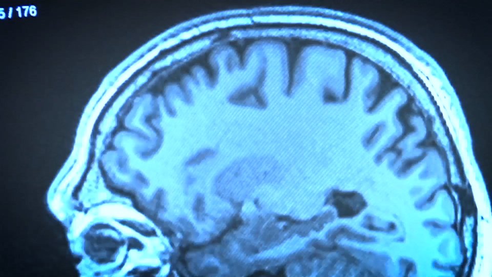
The brain is the most complex organ of the human body. At the very least, it is the most difficult to clarify. However, doctors have gradually invented methods to understand their organization and functions. Without much to clarify, it is surprising what we have learned.
The latest news on this topic comes from the hand of the American neurobiologist Gabriel Kreiman. The Kreiman group has individually analyzed neurons in certain areas of the brain. The participation of each neuron in vision has been analyzed, obtaining surprising results. They find that each neuron is activated by a type of image and not just by specific images. In other words, a neuron does not respond to an apple, but to any of the fruit group. These results will be published in the September issue of the journal Nature Neuroscience.
Behind the confirmation
Similar results were obtained in pot experiments in 1994. Macaques know the faces of their group. Scientists saw how several neurons were activated on any side. However, each face gave a different answer. The macaques, according to these answers, made a concrete face image.
The problem posed by the study of neurobiology is that for ethical reasons very few experiments can be performed in humans. Most research requires the use of animals. But if it is possible to confirm the results in humans a big step forward is taken. Kreiman's research has done so and has proven and extended in humans what they learned with macaques.
He experimented with a group of epilepsy patients. These epileptics did not receive the effect of drugs so they needed surgical treatment. In fact, electrodes were placed in the brain to find out where epilepsy was occurring. The Kreiman team took advantage of the experiment. Seeing images of a topic activated the same group of neurons. For example, images of famous and unknown characters are not treated by the neurons themselves.
Hippocampus
Data were obtained from the temporal branch of the brain. The study has shown that the obligation of the parties is more complex than expected. It is known, for example, that neurons found in the "hippocampus" part have an obligation to become permanent short-term memories. This time the responsibility for the response to the images of spaces (places) has been highlighted. Discovery is even more important, as the hippocampus also treats information for image classification. Keep in mind, however, that epileptic disease has its origin in the brain. Would it have the same results in healthy people?
We are far from knowing how the brain works. However, we gradually understand where each process occurs and what responsibility it has. It is not little.
Buletina
Bidali zure helbide elektronikoa eta jaso asteroko buletina zure sarrera-ontzian


