[Diagnostic imaging]
Decades have passed since the discovery of the possible use of X-rays in medicine; today, many diagnostic techniques allow both to know the functioning of the body and to see the body from the inside. Images are used in many fields of medicine.
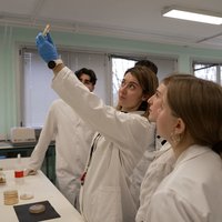
Capturing and interpreting these images is not an easy task. Some research groups at the Public University of Navarra are working on medical imaging. All research efforts are aimed at improving the quality of medical images and facilitating the work of physicians.
More and more technologies are being used for medical imaging: X-ray, MRI, positron emission tomography...
The image is an indispensable tool in medicine. The images visualize situations in which there is not only the possibility of seeing the human body from the inside, but there is no other way to recognize it.
But medical images aren't photographs, they don't show anything that the eyes can see.
ELENA PRIETO; PUBLIC UNIVERSITY OF NAVARRA: Some parameter of a particular organ of the body is represented; these may be anatomical or physiological parameters. Diagnostic information is obtained. Some
research groups at the Public University of Navarra work with medical images. The aim of the studies of these groups is to facilitate the work of doctors.
ELENA PRIETO; PUBLIC UNIVERSITY OF NAVARRA: The objectives are different: access to most of the information and/or the accuracy of the information that
will be extracted. Medical imaging uses a variety of techniques.
ELENA PRIETO; PUBLIC UNIVERSITY OF NAVARRA: Different information is obtained with each technique; the information depends on the type of image. The parameter that is obtained with the type of image that we work with is functional, that is, it measures how the organism works. Other types of imaging, such as anatomical imaging, magnetic resonance imaging, or computerized axial tomography, can measure things related to tissue density or the appearance, morphology, of organs.
Regardless of the technique to be used to acquire the body information, a process known as reconstruction must be performed in order to be able to view the information in the form of an image, i.e., to form the image itself.
ELENA PRIETO; PUBLIC UNIVERSITY OF NAVARRA: The information is acquired in a manner that needs to be reconstructed before the image is acquired. In order to determine the final image, there are several critical parameters and options in the reconstruction process.
Depending on which parameter is used in the reconstruction process, the quality of the image to be acquired will be one way or another.
ELENA PRIETO; PUBLIC UNIVERSITY OF NAVARRA: And even the best image quality available will have its own noise and, in addition, it will not have a good spatial resolution. Therefore, anything that can be done to improve what has been achieved will help to improve the diagnosis by imaging.
In order to improve diagnostic imaging, several studies have been carried out at the Public University of Navarra. To facilitate the diagnosis of brain tumors, image reconstruction has been optimized and magnetic resonance imaging has been used to improve the resolution of images obtained by positron emission tomography.
Research is being carried out not only to improve the quality of images, but also to facilitate decision-making on medical images. The research group, led by Humberto Bustince, uses artificial intelligence systems to facilitate decision-making by physicians.
HUMBERTO BUSTINCE; PUBLIC UNIVERSITY OF NAVARRA: Decision-making systems, that is, systems that represent the knowledge possessed by the expert, to cultivate that knowledge, to cultivate the image itself and to make decisions. What we want is for the system, in this case the computer or the expert, inside the machine, to help us make the right decision.
Initially, these systems were used to diagnose skin cancer.
HUMBERTO BUSTINCE; PUBLIC UNIVERSITY OF NAVARRA:
In any image it is very clear that some pixels or elements of the image are of the background; others are clearly of the stain. But there are also some pixels where it is not possible to distinguish whether they are from the background or from the stain, pixels that are not even distinguishable from those of medical experts. As a result, we have strived to implement the doctor’s expertise into a program.The knowledge is in the databases, but the experience that gives a doctor the ability to know that a stain is dangerous, the experience that he needs to decide to have the operation, is what we have tried to implement.
The Bustince team offers support to the University of Waterloo in Canada for the diagnosis of prostate cancer patients. The systems they have developed help them to distinguish the objects that appear in the images, which facilitates the diagnosis of the doctors.
Ultrasound, nuclear medicine, scans, or MRI are widely used medical procedures, but in the short term of two or three years, millimeter waves will probably also be used to take pictures of the body.
MARIO SOROLLA; UNIVERSIDAD PÚBLICA DE
NAVARRA:Most molecules and tissues of interest to life respond very differently under the influence of this type of light, the millimetric light in the middle of the terahertz. As a result, we expect to see features that cannot be seen with more common techniques, such as X-rays, MRI, etc., which cannot be seen. Therefore, these equipment can be auxiliary tools for medical diagnosis or another option.
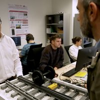
There are very few laboratories in the world that can work with millimeter waves. They have a network analyzer at the Public University of
Navarra. MARIO SOROLLA; PUBLIC UNIVERSITY OF NAVARRA: The equipment allows us to send electromagnetic waves; it is able to send waves with a wavelength of millimeters or less to an object. When the object is moved, the waves pass through it or are reflected in it. With this equipment we will be able to measure the waves that pass through the object, measure how much and how they have passed through and also measure the waves that have been reflected.
Once developed in radio astronomy and the military field, millimeter wave technology can be used in medicine in the short term.
MARIO SOROLLA; PUBLIC UNIVERSITY OF NAVARRA: It will be an additional aid for the diagnosis of situations that cannot be diagnosed with current techniques, especially for the detection of skin lesions. Hospitals in Great Britain and the United States are mainly researching skin cancer and breast cancer. For what reason? Because they are skin lesions that can be seen well with this technique.
Buletina
Bidali zure helbide elektronikoa eta jaso asteroko buletina zure sarrera-ontzian