Images for medicine
At Vicomtech-IK4 they work with medical image processing to develop applications that help doctors in diagnosis and surgery planning.
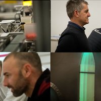
It looks like an operating room, but it's a lab. Even those who wear a white robe are not surgeons, but engineers and computer scientists. We are located in the Vicomtech-IK4 research centre, a department dedicated to the development of health applications.
They focus mainly on the analysis, processing and visualization of medical images for hospitals and hospital providers.
Assisted by SHABS RAJASEKHARAN. More places to stay in Vicomtech-IK4: We work mainly for companies. For example, for suppliers of machines for hospitals. Because they want to add value to these machines, or they want to use intelligent software... We mainly make software.
A software is being developed to control this microscope over the Internet. The main challenge is to transmit high-resolution images without losing quality.
JON PEÑA LEGARRETA. More places to stay in Vicomtech-IK4: In many cases it is the case that in a geographical area there are no experts or professionals properly trained to interpret this image, and the image must be sent elsewhere. With this software, the expert located elsewhere could control the microscope so that he could go to the point of interest and see the image in real time.
This other software serves to visualize medical images in an intuitive way. In this case, we are studying the thorax and abdomen of a patient through tomographic images. What you see on the top left is an image taken by the CT from above. This program shows the same body point laterally and anteriorly, and can also create a more complex fourth view.
JON PEÑA LEGARRETA. More places to stay in Vicomtech-IK4: In the coronal view it is very difficult to see the entire spine. So, through a software process, we trace a line and create a view that follows that line. Somehow, this line is corrected and we can get a complete view of the spinal cord.
The visualization of images in this way is useful for making diagnoses; as well as for planning an operation. Image-guided surgery is based on this idea: images previously extracted from the patient are used as maps by the surgeon.
In order to know where it is located on this map, certain points are marked on the patient's body by means of a punch with sensors. That is, the body is given a series of coordinates to unify the image and the reality.
These images are currently displayed on the monitors of the operating room, but in the future they want to take advantage of augmented reality.
JON PEÑA LEGARRETA. More places to stay in Vicomtech-IK4: Instead of viewing them on monitors, the surgeon could see them in the intraoperative microscope’s eyepieces, superimposed on the image taken by the microscope, where the Willies circle is located, or he could see the arteries, veins... with a different contrast and in a different color, to have a different visual aid.
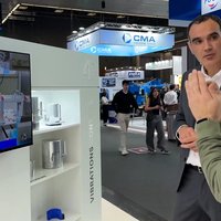
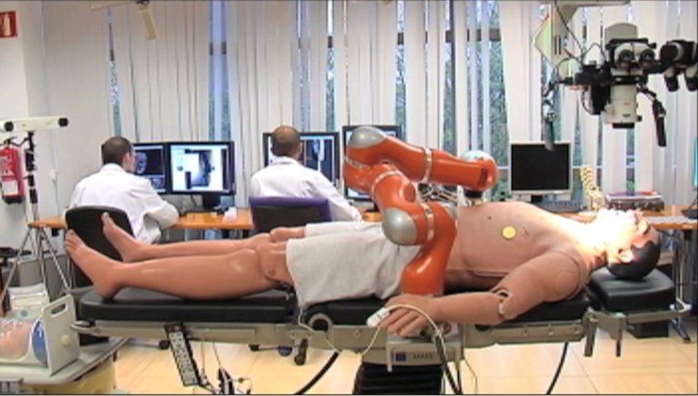
However, acquiring medical images to work on projects is not easy, so they are created in the laboratory itself. For this purpose they have phantoms commonly used in the training of doctors in faculties: a bronchial tree for endoscopies, a doll representing a 21-week fetus, and Patxi, an imaginary patient.
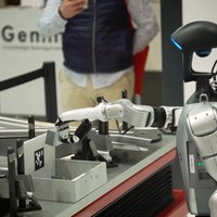
What is moving over Patxi is a robotic arm commonly used in the automotive industry. Like most surgical robots, it can be remotely controlled and various commands can be programmed to operate autonomously. The platform that is being developed here will offer the surgeon something more: a tactile sensation.
JON PEÑA LEGARRETA. More places to stay in Vicomtech-IK4: Robots currently used in surgery, although they are innovative and advanced machines, still do not offer this sensation of contact with tissues. It is not the same as cutting muscle or bone, and the force that is exerted must depend on it. We have managed to have a feedback of the force that the robot exerts on the phantom.
Assisted by CAMILO CORTÉS. More places to stay in Vicomtech-IK4: We installed a force sensor on the robot. We send this information to a haptic device, which exerts its force through a series of actuators.
Assisted by SHABS RAJASEKHARAN. More places to stay in Vicomtech-IK4: Sometimes ideas come out. We talk to doctors (we work with BioDonostia, for example), we explain our ideas and we analyze if they make sense in their field of work. Other times, companies turn to us. They know very clearly what they want, and they come with a specific plan: ‘Look, we have this product and we want to add value to it. What do you offer us?”
[Robotic and image-guided surgery as a means of approaching non-invasive medicine]. The contribution that computer science has made to medicine so far is great and the possibilities it offers for the future are spectacular.
Buletina
Bidali zure helbide elektronikoa eta jaso asteroko buletina zure sarrera-ontzian