The scientific photography
Picturesque images of nature, small to large, things that can only be seen under a microscope, photographs obtained using infrared or ultraviolet light, three-dimensional images... These and many others complement the specialties of scientific photography.
Beñar Kortabarria; Elhuyar Foundation: Today Luis Monje shows us what scientific photography is and how it is done. Luis Monje is one of those great masters of scientific photography who works here at the University of Alcalá.
The Scientific Photography Service is a support center for the university’s medical biology team, led by Monje. Although Luis Monje has hearing problems, we didn’t have any problems understanding each other
Beñar Kortabarria; Elhuyar Foundation: How did you get started on this? Luis
Monje: I was finishing my biology and I was doing my doctorate in botany. The university then took a position as a scientific photographer and cartoonist. I knew almost nothing about photography, but I did a lot of cartoonist work, in newspapers, in comics, and I also did most of the university’s scientific drawings. I introduced myself and began to study scientific photography. But there were no studies, no books, no subjects like this... I was studying for two years until I got a taste.
Beñar Kortabarria; Elhuyar Foundation: It's been a long time since you started. Has it changed much?Luis
Monje: Digital photography has made the image available to anyone. Before it was necessary to play with the diaphragms and until the film was revealed the result was not visible, now with a compact camera it is easy to take photos, even macros. In the past, big shots were needed to take macro shots, while today, with a small camera, it is possible to take spectacular pictures of a fly. Scientific photography has also changed due to the fact that the sensitivity of films and sensors is not comparable; the sensitivity of sensors is much higher. And being able to see the photo as it is taken means saving time, attempts and film.
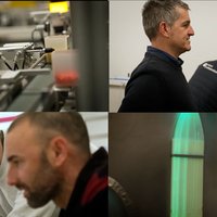
Luis Monje will take pictures of microscopic fungi. The work
starts in the laboratory from the preparation of samples sent from the university’s
botanical department. You have to work by hand and with great care to avoid the breakage of microscopic fungi. Carefully, use the help of both the magnifying glass and the microscope to look for the most beautiful and meaningful view of the samples. In addition to being beautiful, photographs are useful for work.
This preliminary preparation has a lot to do with the result of the work.
Luis the Monk: This requires a lot of technical knowledge. There are special cameras for every specialty of scientific photography, almost tailor-made, but they are very expensive. In one-season pesetas, it would cost more than 7-8 million, and some high-speed cameras reach up to 20 million. So here we take advantage of the old cameras, we do DIY. We dismantle the cameras and adapt them according to the work we have. with cameras of 300 euros we are taking ultraviolet photographs, taking unparalleled images, and with infrared as well. Here, for example, with this material for making macros, we are getting spectacular images with the use of serial cameras and we have reached measures that until now could not be achieved. We replace money with imagination.
These picturesque images are the result of the work mentioned, as they are obtained through a system that has been conceived and executed by Luis Monje himself.
The system is based on a conventional digital reflex camera. Attached to it is a bellows system that allows you to enlarge the image as much as possible. It also has a special lens, very small, that can open the diaphragm up to the size of a needle. To allow light to reach the camera through such a small hole, fiber optic lanterns are used. Since it has a filter for the heat, despite its high intensity, it is a cold light. An image magnification of 20 times can be achieved in this way.
In order to focus the samples at each point, they must be moved. Cut an old microscope to move it, and use its micrometric system.
It's time to take pictures. Fix the sample, focus, and take photos. The focus is shifted from one photo to another by about a millimeter. Then, through a mathematical formula and a computer system, the sum of all the photographs will be made.
Beñar Kortabarria; Elhuyar Foundation:Is this how you get these photos?Luis
Monje: No, I don't. We use an electron microscope.
Beñar Kortabarria; Elhuyar Foundation:So with this system we would get to do things like this and that.
Luis the Monk: We can get here; the limit is there. we can increase it 20-25 times.
Beñar Kortabarria; Elhuyar Foundation: How much weight does scientific photography have in research? how important is it in science?Luis
Monje: The importance is enormous. Keep in mind that research is based on making observable observations: both in physics, taking measurements, and in biology, describing a new species, and in medical interventions... there is a process of observation in all areas. The results are then published, which is very important. Note that a year-long study ends with the publication. Multimedia is becoming increasingly important in science and publications. Therefore, it may happen that they do a huge job and are rejected for having very bad images, or they are not accepted because the images are of poor quality. It can also happen that they have mediocre works, but they are accompanied by spectacular images and they are chosen to be the cover of a magazine.
After taking the photo, it’s time for IT. All captured images are converted into a single photo using commercial software. Thereafter, images such as these can be obtained by using image processing programs.
Buletina
Bidali zure helbide elektronikoa eta jaso asteroko buletina zure sarrera-ontzian