Miraculous images in the light of the microscope
With this photograph of differentiated tumor cells in the brain of a mouse, researchers Bruno Cisterna and Erico Vitriol (Augusta University, USA) have won the first prize in the 50th edition of the Nikon Small World competition.
The cytoskeleton, microtubules and actin nuclei of the cells are clearly visible in the figure. Indeed, Cisterna and Vitriol are investigating how neurodegenerative diseases are affected by disorders in the cytoskeleton of the cell, in particular microtubule disorders. Microtubules function as a transport network in cells, and the Cisterna group has shown that the PFN1 protein plays a key role in the functioning of this transport network (the study was published in the Journal of Cell Biology).
To obtain the image, it took about three months to improve the staining process of the cells, so that all structures were well visible. After the cells were allowed to differentiate for five days, they were observed under the microscope for three hours until the appropriate site and time were found at which the differentiated and non-differentiated cells interacted.
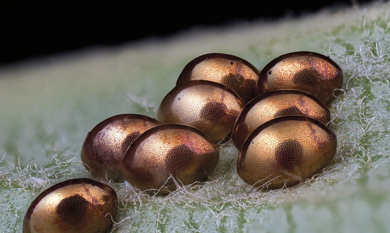
This image has won, but also highlights another 87 of the thousands of images received by the competition jury.
Buletina
Bidali zure helbide elektronikoa eta jaso asteroko buletina zure sarrera-ontzian


