


Chitridiomycosis: A disease that causes panic
The cause of the disease of chitydiomycosis of amphibians is the fungus Batrachochytrium dendrobatidis. This species belongs to the group called Kitridio, Chytridiales order. These fungi are characterized by being a mobile with flagellate spore or zoospora that separates them from the rest of the kingdom of Fungi.
Kitrides participate in the microbial community, both in aquatic and terrestrial ecosystems. In them they can be saprofitos (feeding on dead organic matter) or parasites of microscopic organisms (pollen, algae, invertebrates), which play a fundamental ecological role by participating in the decomposition of sustainable matter (chittin, keratin and cellulose of difficult decomposition). B. B. Dendrobatidis was described in 1998 as the cause of the reduction of amphibian populations. Known by experts, it is the only dew that contaminates vertebrates.
B. B. The ability to infect dendrobatidis vertebrates is based, among other things, on the ability to use keratin as a food. In fact, the surface of amphibians is a thick outer layer of keratin. Being a complex polymer, keratin is not useful for most fungi, since in general they are fed by simple compounds. But the zoospora of this kitridio comes the keratin perfectly. When finding an appropriate host, synthesize and eject proteolytic enzymes and sterases. These enzymes can degrade keratin and drill the surface of amphibian.
As it is a simple fungus, B. dendrobatidis does not develop breeding macroscopic structures (like some complex fungi develop seta structures), so it passes unnoticed at first sight. When it is in the guest, however, it forms a simple body similar to the fondeo roots that are called rhizomizelium. This will fulfill vegetative functions and produce reproducing structures.
Life cycle
As has already been indicated, the infection occurs by the mobile spore. The zoospora is free in the water and is directed to the hostelero. It can swim 24 hours, but its mobility is limited and can not travel long distances. To deal with this limitation and find the guest as soon as possible, the zoospo is used for chemotaxi, that is, its movement is governed by chemicals in the area. Once the host is located, the amphibian, adheres to the outer layer of the keratinized epidermis. There it reabsorbs the scourge, moves towards the bottom of the skin and creates a structure in the form of a pouch: zoosporangio, inside which the reproduction begins. It reaches 4 days and releases about 300 zoospores in the middle. To release the zoospores to the outside medium forms a tube that crosses the surface of the amphibian. The cycle begins again when zoospora finds the right substrate. This substrate can be both the hostelero itself and the other. In this way the amplification of the disease is achieved, both increasing what a hostelero already had, and extending it in the environment.
Pathogenesis and avoidance mechanisms
To understand and stop the disease, in addition to the life cycle and fungus structure, it is very important to analyze the infectious mechanism of amphibian and its effects. We cannot forget that some amphibians have developed a number of resources to deal with this disease that can also help find a solution for the future.
The fungus grows in the outer layers of the keratinized surface of adult amphibians, in the stratum corneum and granular. These layers, especially the corneal strata, have a width of between 2 and 5 ?m. In contaminated animals, on the contrary, this layer is thickened (hyperkeratosis) until reaching 60 ?m. Then, this layer becomes irregular and subepithelial cells explode to make room for zoosporangios. In addition, the release tube of zoospores that each zoosporangio crosses the surface of the amphibian to the outside medium and breaks the skin. This action can be seen microscopically. Macroscopically only late symptoms such as swelling of the skin, redness, increased viscera, bleeding, etc.
Amphibians die between 10 and 18 days after the disease, since their hearts are left. This is due to the importance of the skin in the respiration of amphibians and in the balance of electrolytes (osmotic inner balance). In fact, kitrides mainly infect highly vascularized areas of the skin. This will make it difficult to exchange gases, preventing skin breathing. In addition, the swelling and deformation of the skin causes the interruption of the transport by surface of the ions sodium, potassium and chloride, putting upside the osmoregulation of the animal. As a result, the imbalance of the electrolytes and the decrease in oxygen occurs in the blood, coinciding with the increase of carbon dioxide, which partly chokes the animal and stops the heart.
However, some amphibians have natural prevention mechanisms against the disease. For example, it has been shown that the arrabio Plethodon cinereus and the mucous Rana contain in their skin a bacterium ( Janhinobacterium lividum ) that prevents the development of the fungus. This bacterium releases fungicide substances to the surface and prevents infection.
On the other hand, it has been observed that superficial peptides play a fundamental role against the disease: breaking the membrane of the fungus inhibits the contamination of the pathogen and its growth. Therefore, they avoid the post-consolidation growth of zoospores. However, not all amphibians have this type of defense mechanisms, which has caused the global spread of the disease.
All over the world
As has already been indicated, although some species of amphibians have mechanisms to fight the fungus, it has been introduced very violently in some ecosystems, especially in areas of vulnerability of amphibians. Therefore, chitridiomycosis is currently a widespread disease in the whole world and in all non-Asian continents, amphibians infected with chitridiomycosis can be found. In addition, it must be taken into account that there are large areas that have not yet been studied.
In Europe there is not much information about the spread of the fungus. In the investigations carried out the disease has been detected in six countries of the continent, being three the most affected amphibian species: the txantxikua ( Alytes obstetricans ), the common arrabio ( Salamandra salamandra ) and the common toad ( Bufo bufo ). Most of the cases of kitridiomycosis have occurred in Switzerland and Spain, the latter being the first country that suffered the disease, being in 1997 the first amphibian died in the Natural Park of Peñalara (Madrid). In the summer months of 1997, 1998 and 1999, very high mortality rates were observed in the populations of brooches.
Dissemination and justification of success
Although the life cycle of chitridium, as well as its approximate geographical distribution, are known, there have been problems to explain the widespread spread of the fungus and the current amphibious mortality. Several hypotheses have been formulated.
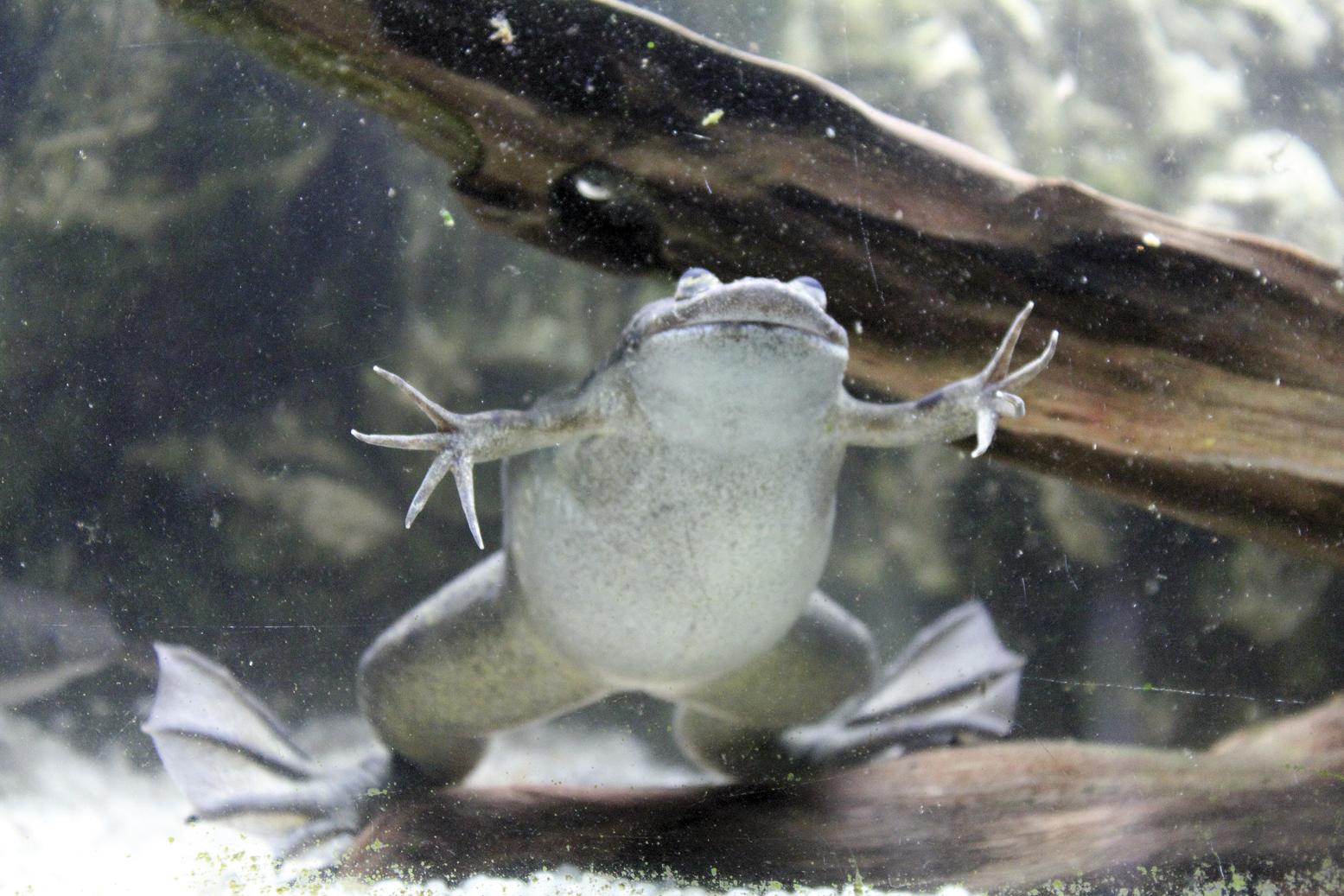
At present the hypothesis has been imposed that human participation seems to be in the African origin of the fungus and that the species of frog Xenopus laevis has had a great influence on its expansion. The man has spread this frog (and the fungus) all over the world, as the research of the pregnancy test. It is believed that this frog has acted as an asymptomatic vector.
In addition, it has been shown that B. dendrobatidis has survival capacity in moist lands and bird feathers. This means that it not only uses amphibians as vectors, and it is believed that when there is a shortage of them it may be due to movements of birds or lands (transported by animals by drinking water in fish sampling, at the foot of man, tires, etc.) and that can survive.
It highlights its different impact on the different species and on the separate populations of the same species. In fact, chitridium has been able to withstand harmful conditions for other organisms, among others. In fact, it maintains the capacity to increase and expand the temperature (4-28 °C) and pH in wide ranges, with the capacity to generate cysts. Therefore, in situations not optimal for amphibians, that is, both at low temperatures and situations of pollution, chitridium will act as opportunistic pathogen taking advantage of the defenselessness of amphibians in these situations.
Although these characteristics are known, there is currently no effective way to fight the disease. Some trials have been conducted at the local level (inoculations of the bacterium J. lividum, based on the intrinsic immunity of amphibians; varying the ambient temperature and pH...), but they are not enough to end the global crisis.
Man also expands
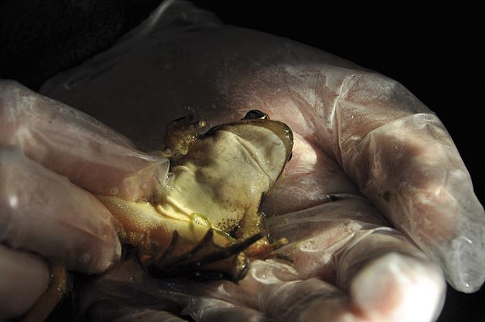
As mentioned, human influence is also perceived in this ecological problem. The main global dispersion has been produced through the species Xenopus laevis, but the influence of experts on amphibian research has also been verified, to the extent that field work is an important part of the research of amphibians and disease. The researchers move from one humid zone to another in which amphibians are found and, consequently, they behave accidentally as dispersion factors at a local scale. For this reason, there are a series of preventive measures to take into account, such as the use of disposable gloves for the collection of each amphibian specimen, the disinfection of all the material before its reuse once the field work is finished, the prior sterilization of all the material to be used, the extraction of specimens killed by disease.
Human activity has caused a great impact on the group of amphibians and remains so. It is estimated that the disease has eliminated about 200 species in the last 30 years. How far will we continue without taking care of our surroundings? What is our limit? Unanswered questions, for the moment...
References References References References
Buletina
Bidali zure helbide elektronikoa eta jaso asteroko buletina zure sarrera-ontzian


