
Interior photography with ultrasound
2008/01/01 Kortabitarte Egiguren, Irati - Elhuyar Zientzia Iturria: Elhuyar aldizkaria
Ultrasounds have been known for a long time. These are sounds of more than 20,000 hertz, so the human ear cannot perceive them. The behavior of the ultrasound is the same as that of the sounds we hear: means are used to move from one medium to another --air, human tissues..-, move at a certain speed, have waveform and transport energy.
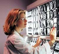
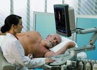
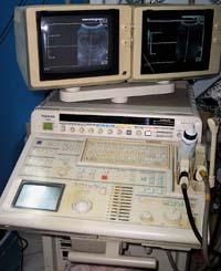
These sounds have many applications. Being able to access the inside of the materials and get a lot of information about it, in many sectors the ultrasounds are a useful technological tool. But perhaps one of the best known uses is the study of biological tissues. As mentioned above, ultrasound scans are based on ultrasound. For the realization of the ultrasound, first the part of the body to study is moistened with a gelatinous substance. This gel works as a driver. A transducer is used for sending ultrasonic waves. The transducer is used as sonar and directs the waves toward the patient. The sound of the transducer is reflected in the structures inside the body, and a computer analyzes the information of those sounds and creates an image on the screen.
In short, the ultrasounds are based on the physical principles of radar that use bats or ships; that is, the echo that occurs when colliding the sound waves against a tissue indicates its position, size and composition (solid, liquid, mixture).
Not only women but men
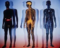
Ultrasound scans are usually done to look at the internal organs and blood vessels: belly, breasts, pelvis, prostate, scrotum, and thyroid, among others. Although ultrasounds are often associated with women, they are also performed by men.
Abdominal ultrasound, for example, can detect tumors of the liver, gallbladder, and pancreas in both men and women, as well as intraabdominal tumors. It is usually used for the study of kidneys, liver, pancreas, spleen, and abdominal blood vessels. It is very useful to diagnose causes of abdominal pain such as appendicitis or kidney stones or gallbladder. In fact, it informs us of the size of the abdominal organs, it indicates whether or not there are remains of tumor in them and the origin of them. It also serves to investigate jaundice, as it allows the diagnosis of the dilation of the bile duct.
Abdominal ultrasounds are used not only for the diagnosis of the aforementioned diseases, but also to guide operational processes such as needle punctures of abdominal lesions (which serve to obtain histological diagnosis or microbiological analysis) and abscesses, as well as for the implantation of drainage catheters of groups of liquids.

Breast ultrasound scans are usually done to women. They are used to differentiate tangible nodules that can appear in mammograms and/or in the gynecologist. The main objective of this type of ultrasound is to detect if the tumor is solid or liquid to study its goodness. It offers very good images, except in very obese women. As mentioned above, the main goal is to identify whether the nodule or tumor that has previously been observed by the doctor in contact with it or through mammography is either solid or liquid.
It is not a substitute technique for mammography, but complementary. In addition, in general, once the mammography has been performed, radiologists also perform an ultrasound to further deepen the diagnosis. The separation technique of solid and liquid structures is especially suitable. Therefore, it is often used in the detection of chest cysts, small structures filled with liquid.
For the diagnosis of prostate cancer in men, rectal ultrasound is performed. For this purpose, a probe of approximate size of a finger that emits ultrasound is inserted through the rectum. These ultrasounds cause echoes when colliding with the prostate. All these echoes are again collected by the probe and processed on the computer until the image of the prostate is generated on the screen.

When the catheter enters the rectum it is possible that the patient receives some pressure. The process lasts for a few minutes. Likewise, rectal ultrasound is one of the most commonly used methods for biopsy. In fact, prostate tumors and normal prostate tissue, in general, reflect different sound waves. Therefore, rectal ultrasound is used to guide the biopsy needle to the exact location of the prostate tumor. However, rectal ultrasound is not recommended in cases of early diagnosis of prostate cancer.
Follow-up to pregnancy
In general, ultrasounds do not produce wounds, do not produce pain, and are fast techniques. In addition, no radiations are used, although they are performed in the radiodiagnostic service. For this reason, eleven diseases are used not only for diagnosis but also for fetal development during pregnancy. In fact, ultrasound scans are a safe interior observation system, according to the research carried out, it does not appear that the ultrasounds damage.
The use of ultrasound in the field of obstetrics began in 1958, being one of the most memorable episodes of medicine. In fact, for the first time a safe examination of the fetus and its surroundings (placenta, amniotic fluid, etc. ). Over the years, its applications have spread not only to the field of diagnosis, but also to other areas.
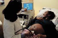
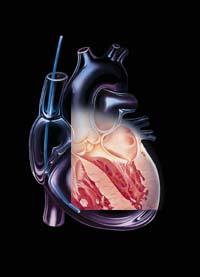
Another application of three-dimensional ultrasound in obstetrics is the detection of fetal anomalies. Facial, limb and spine lesions can be observed, as well as their possible use in the face of heart problems. This technique detects no more malformations than two-dimensional ultrasound, but shows with greater precision the severity of these lesions. However, according to Juan Cruz Trecet, a gynecologist at the Donostia Hospital and Polyclinic Gipuzkoa, 99% of the ultrasound scans currently being carried out are still performed in two dimensions.
When three-dimensional images are added movement, they are called four-dimensional ultrasound. These ultrasounds allow to study in real time a certain fetal behavior. In fact, they observe the movements of the body and face of the fetus. With this technique, fetal malformations can be observed in greater detail as abnormalities of the legs and hands, spine bifida and tumors.
In short, it can be said that it contributes to a more accurate diagnosis of the fetus. In fact, observation of face movements often helps experts to obtain additional information about the fetus. Moreover, the gynecologist Juan Cruz Trecet comments that it is more a matter of visibility. In your opinion, 4D ultrasounds would be adequate to resolve doubts that may arise in 2D ultrasounds.
Whatever the ultrasound of two, three or four dimensions, there is no doubt that this technology tells us of non-emerging diseases, and not always of diseases, which will continue to give.

Gai honi buruzko eduki gehiago
Elhuyarrek garatutako teknologia