
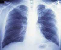
X-ray images at lower doses
2002/05/20 Lasa Iglesias, Aitziber - STEAM Hezkuntza arloko arduraduna
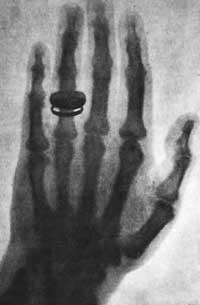
Conventional x-rays show the "shadows" of the tissues. That is, it emits x-rays and collects them a special photo plate, but if we place the arm in the center, since the tissues and bones of this arm absorb light, and not all in the same way, in the photo appear different shapes: bones, muscles, etc.
But the newly developed technique is based on the refractive index of light. Inventors have called it phase contrast radiology. When light strikes on a material, some rays are reflected and others pass through the material. The latter are called refracted rays and depending on the material traversed, the angle that forms the attack ray with the surface is different. The ratio is given by the refractive index. If you can know that angle, you can form the image.
The advantage of this new technique is that you can work at much lower doses. It seems that it will give much more information than conventional x-rays. For example, the new technique will detect tumors from the beginning.
However, the need for highly collimated rays prevents the use of x-ray machines that have so far been used in hospitals.

Gai honi buruzko eduki gehiago
Elhuyarrek garatutako teknologia