


X-rays
The X-ray is an invisible light and has a wavelength ten thousand times shorter than the visible light. Electromagnetic radiation known as X-rays is emitted by cathodic tubes.
Explaining what X-rays are is nothing difficult at present, since they are well known. But a century ago those rays of light were not even known. We owe a German to “find” X-rays with application in different fields.
It has always been said that discoveries are achieved thanks to work, knowledge and time, but in some cases luck also helps a lot. In 1895 it can be thought that in the scientist of Wilhelm Röntgen all these factors converged, working in the laboratory of the University of Babiera to cross the electric current.
A mysterious ray
One afternoon, however, something special happened in the laboratory. While working with the essay, the bottle in a balda was clarified. In the bottle was found fluoro, specifically barium cyanide. The German professor was surprised, from the session he was investigating came the light and those rays were the ones that illuminated the bottle. Those unknown rays did not cross the cyanide barium and that is why the bottle was lit. In view of this, Röntgen began to investigate materials that could be traversed by the rays. To begin with, he placed a piece of cardboard at the height of the essay and when the light was done, the beams illuminated the bottle of the balda.
Seeing that the light was passing through the cardboard, next to the essay, he placed as a screen a piece of paper rubbed with the cyanide. The paper absorbed the mysterious rays taken from the session and the screen was illuminated. Therefore, these unknown rays could cross the cardboard, but not the barium cyanide screen. While conducting tests of the absorption of light by lead, Röntgen had a great surprise. He grabbed the lead between the program and the fluorescent screen. The lead absorbed the light and in the fluorescent screen appeared the shade of the lead.
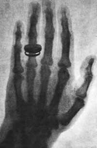
However, in addition to the shadow of the lead, also appeared the shadow of the fingers of the hand that grabbed it; more specifically, the bones of the fingers were ‘reflected’ on the screen. The shadow of the bones appeared clearly on the fluorescent screen and the hand meat could also be highlighted, although more diffuse than the bones.
The whistle of the bones of the hand that appeared on the screen is called radiography today and, as we have seen, it was the first time it was achieved by shooting X-rays of the essay. Also curious is the name that the German put to lightning. Mathematicians call X to the indeterminate amount and following that “custom” Röntgen baptized the ray with the name of X-rays.
Use of X-rays
X-rays explore the object placed on par and the shadow of the object is collected on the fluorescent screen or in the photographic emulsion.
When we talk about x-ray scans, the ones that make us in the hospital or in the outpatient immediately come to mind. However, we should not forget that in addition to exploring our internal organs and bones, X-rays are very useful for many other tasks.
In medicine, various techniques are used to perform x-rays. The illusions of the organs and bones of the body are recorded in the phototype or photographic emulsion, but all the shadows are not seen with the same clarity and precision. For example, the x-rays of our skeleton are highly accurate; metallic objects also absorb X-rays very well, and more than one time metal objects have been found in the internal organs of the patients. On the contrary, organs such as the bowel, urinary system or veins are not so “clean” and to achieve proper precision, the patient is made to take chemicals.
In addition to in medicine, X-ray is also widely used in the industry. X-rays are made to correct changes in chemical compositions and “errors” that occur inside the materials. In the foundational laboratories, for example, the quality of the welds is controlled by X-ray.
Another important use of radiography is given in the artistic works and, above all, in the paintings. X-rays allow to analyze the state in which the paintings are found; in addition to studying their conservation status, x-rays allow to know the age of the painting. Radiography also “denounces” the techniques used by the painter or the last touches he has given to the work.
A hundred years ago, Röntgen discovered X-rays and, as with all discoveries, the X-ray technique has been improving over time. When we talk about supports, we have mentioned fluorescent screens and photographic emulsions, but currently the computer supports have been advanced. Now the silk (digital x-ray) of the explored object is stored on the computer. The use of computer supports offers great advantages. On the one hand, the information (radiography) is stored in computer support with greater security and can accumulate more information. On the other hand, we will be able to treat the illusion that will appear on the computer screen; expanding or reducing the image, we will be able to accurately analyze the desired point.
Buletina
Bidali zure helbide elektronikoa eta jaso asteroko buletina zure sarrera-ontzian


