


Evolution of surgical techniques in cataract operation. Placement of artificial crystalline
The opaque, total or partial, diffuse cataract of the lens of the eyeball is called. The empirical attitude towards the eye attacks that could be detected with a direct observation allowed to warn that the pupil of some of the people who suffered a decrease or loss of vision had a gray or totally whitish hue, while in normal people it was black.
Although this event was somehow clarified, the following theory was formulated: it would be a liquid flowing through the brain; through the visual nerves (which were considered empty tubes) it was deposited in an absolutely theoretical space between the lens and the pupil. The Greeks called it hypochine, sufusions and soups and the Arabs summarized in one sentence the patogeni mechanism and the specific designation.
This phrase was taken to the School of Salerno by a traveling friar who has passed into history with the name of Constantine African. This phrase became too long and uncomfortable to designate the diagnosis and taking the idea of water falling in one place, it was summed up in one word: cataract or water fall. Subsequently, this term passed to almost all languages, although in the languages of Latin origin there is a certain difficulty, since it is not easy to think that inside the eyeball can produce authentic water slides.
In Basque, however, the people have designated this situation by another means. Seeing that the eye loses its transparency, that is what the Basques have wanted to highlight. The best known expressions would be BEGI-LAUSOA or BEGI-GANDUA.
Cataract, therefore, would result from brain disease and visual nerves. The corrupt mood resulting from the disintegration of the visual spirit stood in the gap between the pupil and the lens, partially affecting the lens. The chaos of the aqueous and the pneum that was produced in this space destroyed the sight, so it was assumed that the opaque element was partly in aqueous (aqueous) humor and partly in the lens.
The elimination of this obstacle located in the pupil by means of a well-systematized surgical intervention and with a naturalistic technical criterion arises for the first time in the bloated medicine. His route in the writings of the surgeon of Benares (B.C. IX. and VIII. centuries) describes in detail the operation of cataracts and the hygienic and asepsive standards to maintain.
Much later, XII. In the 19th century, the Cordovan oculist Al-Gafiki described the operation of cataracts by reclining the lens, following almost completely what Susluta has told him.
Already in 1583, Bartisautza described the natural absorption of a piece of crystalline in the antechamber and from here came to propose a new technique that was effective in certain types of cataracts.

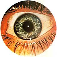
4. Cornea camera
5. Pre(ko)
6. Pupil
7. Crystalline
8. Irisa
9. Vitreous humor
10. Sclera
11. Coroides
12. Retina or bed
14. Visual disc
15. Visual Nerve
16. Ciliary body
17. Camera AStze
18. Suspensive lens ligament (eyelash zone)
Jacques Daviel (1752) has the merit of extending extractive conditions to almost all types of cataracts. This same author obtained a great success of intracapsula extraction of cataract h.d. especially the lens, when he did it with tweezers. This procedure (with variations --clamps, suction cup, freezing of crystalline cryoextraction, etc.-) has been the alternative technique XX. throughout the century.
Among these surgical procedures stands out the participation of the Spanish, proof of this is the prestige and prestige achieved by doctors Hermenegildo Arruga, Ramón Castroviejo, Ignacio Berraquer and his son José Ignacio.
Extraction of the opacified lens allowed patients to regain sight almost immediately. However, the vision, except in the case of certain myoperas, was very sketched, so they were in almost all impossibility of carrying out many activities, which meant a great degree of dependence or dependence.
To compensate for the loss of vision caused by the extraction of the lens, it was necessary to use corrective crystals with adequate dioptric power (mounted in an antioju), which allowed to achieve a visual acuity of ten decimals, both in the near and distant vision. However, the reduction in the patient's rhythm and the insecurity in the perception of the medium as a result of the spherical aberration of the lens conditioned the normal course and therefore the patient's independence.
When the intervention was performed in one eye, binocular or visual vision was impossible, since the dimension of the images received was very different to allow a correct and adequate fusion. This implied a lasting double vision and in many cases forced an eye to a stale ring.
The use of contact lenses with an increasingly precise and fine processing has allowed to solve most of these problems, and since the contact lenses adapt to each specific case, it has been the first time that it has been possible to obtain an optimal visual vision of the eye and a greater field width and visual acuity without distortion of the images.
We want to remember that contact lenses are older than current propaganda can assume. The first appeared in 1887, made by Eugene Pick of Zurich, and in case of afaquia (h.d. in the absence of crystalline) is considered especially useful. An optical of Paris, E. A known as Kait, he also made contact lenses in 1888; floating lenses (loose, loose) of about 8 mm in diameter.
Since then the evolution of contact lenses has been linked to the research of new materials. In this way they have been able to achieve lighter lenses, permeable to gases, progressively improving their transparency or transparency and allowing greater tolerance to patients. The long duration has facilitated the resolution of many optical and therapeutic problems. But as often happens with the appearance of new elements, new lenses have caused other problems, since the wearer of these lenses must maintain careful control and hygiene, with regular medical control and surveillance.

The history of intraocular lenses has been long and fascinating in many fields. Today it is impossible to decide who and how it occurred to him to replace with an artificial lens the opaque lens for the first time, but despite being surprised by the famous knight Casanova in his prestigious Memories, already tells us a curious talk with a traveling ophthalmologist named Tadini in 1766.
This tadini asked him to steal from a Duchess to obtain operating permission to replace his lens in situ with a glass lens. It seems that the operation was not carried out, but nevertheless, an ophthalmologist from Dresden, who named Tadini as a novel, carried out the same operation proposed by him in the city of Leipzig and in 1797, with fatal and predictable consequences, according to the Swiss oculist Schieferli.
After a long silence, J. In 1940 Föster published a humour article in the Leeds Medical Society Magazine entitled An oculist in America. In this work, Föster faces the pros and cons of the procedure from a bold, curious and analysing vision of a young America and its rich economic environment.
Observations made by the English ophthalmologist Harold Ridley: A relatively large number of RAFS pilots, with penetrating wounds in the eyes, supported without discomfort the parts of the abonic garling formed by methyl-methacrylate inside the eye. In 1947, during a surgical session, a student asked Ridley a seemingly obvious question: - Sir, after removing the opacified lens, don't you put anything in place afterwards?
Comparing the authority of the questionnaire to the level of the question, the response was immediate, indicating and justifying that, as usual, optical compensation was obtained using appropriate antioju. But Ridley's clear intelligence did not discard the suggestion and from there a working theory was formulated and launched that opened a new and fundamental section in the history of the cataract.
Taking the human lens as a model and with the help of Mr. Ernest Ford of Raynords Optical, Ridley designed and prepared a lens composed of Perspex CQ. This topic was only the methyl-methacrylate polymer manufactured by the Imperial Chemical Company.
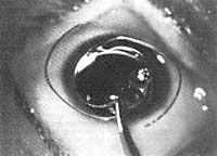
After having a supposedly correct and adequate lens, Ridley performed an extraencapsulated cataract extraction, h.d. the removal of the masses that constitute the thickness of the lens, respecting the posterior capsule as a point of support or as a plane of containment, and the subsequent placement of a new artificial lens in this previously cleaned space.
The lenses initially used were too heavy and because they did not have proper fixation, there were always complications before or after and occasionally they ended dramatically.

In 1964, after fifteen years of constant research on this subject, Ridley operated 70 cases on his own. Hirschman (1984) states that for 25 years he has had the opportunity to analyze a patient who has carried a Ridley lens, with a high tolerance, and who has reached a vision of seven tenths.
The critical analysis of the procedure, but at the same time objective, made clear that it was possible to establish a well-systematized surgical procedure: the extraencapsulated extraction of the cataract and the subsequent placement of the intraocular lens, being indispensable condition to reduce previously the weight of the lens, improve its design and achieve an effective and safe sterilization.
Another problem that highlighted the experience gained over time was the opacification of the rear capsule. In these cases, both before and now, discipleship (or separation) of the posterior capsule is essential. This DISCIPLESHIP, which once required a new surgical intervention, can be solved in most cases in a simple, comfortable and simple way, using modern YAG lasers.
Due to the problems posed by the rear camera lenses and the complications arising from their location and containment system, some surgeons devised new placement and fixation systems to prevent the mooring of the lens, so that, while still placed in its proper place, it was accessible for ophthalmologist control. This new trend opened the era of the intraocular lenses of the pre-chamber.
It was Italian professor Benedetto Strampelli who first designed the lens that was only pre-camera. In 1953 he described a meniscal-shaped implant (radius about 12 mm and rectangular shape). In the central space or gap the optical lens was located and the rest of the implant corresponded to the supporting structure. At the same time, in London, Peter Choyce also chose to perform and position the implant in the pre-chamber, designing the lenses known as Mark VIII and IX.
The advantages of implantation or implantation in the pre-chamber (i.e. before the pupil) can be summarized in:
- In its entirety it is of a single piece and is formed by a single material.
- Its correct and correct centration does not cause alterations in the dynamics and/or position of the pupil.
Its consolidation is firm, without possibility of displacement.
Being a monoplanar structure, the distortion applied to the wound is minimal.
It can be used after sectorial extraction of the iris. It can be implanted in an eye previously
operated with cataract by intra-capsule or extra-capsule procedures, or even as a secondary application.
The disadvantages of the procedure would be:
- The models described had a particular size. Therefore it was necessary to have at least three lenses in each case. (Currently this difficulty is fully resolved with pre-camera lenses with elastic handles).
- The incarceration or retention of angles that caused pupillary distortion was frequent.
It was difficult to achieve a complete adjustment, since changes in eyeball dimensions could not be measured in half a millimeter. (Current lenses have overcome this difficulty.)
The implant more easily endured trauma. Ubeitis,
glaucoma and hiphema syndrome could be conceived as one thing, to the point that, in many cases, the application with severe visual alterations required a new removal.
The changes in the design of these implants have been very numerous, taking into account their location and their points of support and fixation. Some authors used iris as support, fixing it on it, such as those designed by Epstein in 1953-59. Clip lenses were also used in the iris: Developed by Binkhorst in 1958, with modifications of Fiodorov or even of Worst (the latter joined the lens to the iris to keep it thus established).
This wide and different experience showed that capsule fixation had advantages over iris fixation. Iridodonesia and facodonesia (implant displacements and unconventional iris mobility) disappeared almost completely. Binkhorst, Simcoe, Schearing (modified from Barraquer), etc. J loop lenses were highly effective, while presenting the advantage of good sustainability.
Kratz, Charleux, Quintana, Menezo and many others improved their designs and today there is a wide variety of quality visual implants.
The choice of Perfes with visual implant therefore requires a personal experience in relation to the surgeon and a critical and objective opinion that allows selecting the most suitable lenses in each case.
Aware of the importance of the topic and the need to update and exchange information and experience, the Department of Ophthalmology of the University of the Basque Country organized on July 4 and 5, 1986 a course on artificial crystalline surgery and on February 27 and 28, 1987, the III Spanish Club of Nonrefractive Ocular Surgery. Meeting.
The academic, practical, clinical and surgical sessions took place at Policlínica Gipuzkoa. This course was attended by General Optika-Domilens and the meetings of CECOIR last February of more than ten pharmaceutical laboratories, manufacturers of intraocular lenses, etc... The surgical sessions were recorded in a high-quality video tape for later distribution to the different hospitals and university centers.
In these courses the current problems of visual implantation of the lenses were addressed and in the talks and round tables the following teachers and doctors participated: Barahona (Salamanca), Garcia-Alix (Madrid), Coret (Badalona), Menezo (Valencia), Quintana (Barcelona) and Sayans (Plasencia).
Live surgical sessions were also held, with the help of Doctors Menezo and Quintana and Professors Cherleux from Lyon, which were completed with interviews between the attendees. In both acts the immediate translation was made by Dr. Laura Munoa.
In the meeting of CECOIR, the topic of Retinal Emission in the psedophagus was discussed, through a keynote speech, Dr. Ducorneau of the Soudille Hospital in Nantes and Dr. Quentel of the Ophthalmology section of Professor Coscas of the University of Paris, responsible for the theme of the YAG Laser Application, giving a theoretical course.
Different themes were played in the round tables: The multisification of facoes moderated by Dr. Sayans, the design and possibility of intraocular lenses by the hand of Dr. Quintana and the implantation of small power intraocular lenses in high myopia, led by Dr. Menezo. The meeting was enriched and completed with the loose communications of other doctors.
Buletina
Bidali zure helbide elektronikoa eta jaso asteroko buletina zure sarrera-ontzian


