
Form the three-dimensional atlas of the mouse
2001/04/11 Galarraga Aiestaran, Ana - Elhuyar Zientzia
So far, researchers with mice had no reference. Due to increasing knowledge of mammalian genes, along with knowledge of how and where genes act, a localization model was needed.
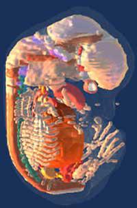
MRI images are widely used to obtain images of human organs and soft tissues, and this time they have been used to study the planes of tiny mouse embryos. These images have been placed in a three-dimensional digital model and have formed an embryo visible from any angle. Soon, analyzing every day the growth of the embryo, they plan to make a map in four dimensions. In this way, you can visualize the mouse development in space and time.
In traditional technique the embryos are cut into fillets and taken pictures. The imaging technique used has prevented the tilting and distortion of the tissues. However, it is inconvenient that intracellular structures are not visible.
The next challenge is for scientists to put data on gene manifestations in the atlas. In this way, an increasingly complete database would be obtained. Other biological data can be added to the map, such as nerve connections and cell migration models.
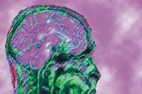
On the other hand, projects are underway to complete maps of other developing agencies. The digital map of the human brain has already been developed and it has been proposed to make fly and chicken.

Gai honi buruzko eduki gehiago
Elhuyarrek garatutako teknologia