

PET, excellent and expensive
2001/03/21 Carton Virto, Eider - Elhuyar Zientzia
Limit of cyclotrons
Positrons are subatomic particles, equivalent to electrons but with positive load. In oncology, the PET technique consists of the use of positron emitting radioactive substances to locate the tumors. The process is done in two steps, first creating radioactive tracers and after injecting the tractors to the patient.
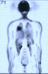
Tractors are an organic molecule adapted to be radioactive. These molecules have a biological function and participate in physical-chemical phenomena that take place in tumors. In addition to detecting the location of the tumor, it is possible to detect and quantify many physiological, metabolic and biomolecular changes that occur in cells. Dr. Arrate Plazaola of the San Sebastian Oncological Hospital considers an extraordinary technique in the detection of tumors. "PET allows to detect very small changes and with a single test you can see the whole body," explained Dr. Plazaola, "which are great advantages." PET is the most suitable technique for locating tumors, but it has a great limit: the cost.
The technique was developed in the 1970s and has been widely used in biomedical research, but the need for cyclotrons for the production of tracers has greatly limited its extension. And it is that the cyclotrons are very expensive. On the other hand, most of the radioactive markers disintegrate very quickly and there was no choice but to use them at the birthplace.
Long life tracer
The fluoro-deoxi-glucose-f18 (FDG) tracer, however, has an average life of 110 minutes and can be transported from one place to another. It is the only tracer of these characteristics that allows to perform the PET in those centers that currently do not have cyclotron. It is enough to have a device that collects the signals issued by the tractors.
In the Basque Country there are two hospitals, one in Bilbao and the other in Pamplona. In Navarra they have the complete apparatus, that is, the cyclotron and the experimental chamber, while in Bilbao they only have the camera and the tracers are carried from outside. If necessary, other hospitals ask for testing. However, Arrate Plazaola tells us that PET is only required in very specific cases: "When there is a cancer focus and we do not know if there is more, when metastasis has been found but it has begun or not, when we do not know, or when after a treatment we have left the body completely clean.
Arrate Plazaola has confessed that if we had it, we would probably use it more times. With the aim of simplifying and embracing the PET technique, there are numerous research underway around the world. The latest news comes from the University of Glasgow, where it is trying to develop a PET appliance that can be desktop. The research has been released today at the congress of the British Institute of Physics.
The device has been made from an optical technique developed by nuclear physicists last year and works with compressed and amplified laser pulses. The pulse is emitted against a sheet of uranium, causing the fission of uranium and releasing particles of great energy. According to the researchers, it can be an apparatus capable of replacing cyclotrons and the first prototype is already being prepared. For the moment they want to apply PET because they know how to design a laser to make PET, but they believe that in the future it will be possible to adapt it to treatments with protons. And it is that the protons have more and more repercussions in oncology.
Less lateral damage
Electrons and gamma rays are currently used to remove tumors by radiation therapy. The rays are emitted as localized as possible, but it is impossible not to affect the adjacent healthy tissues of the tumor, or the tissues that are passed into the tumor. Damage can be especially serious in brain tumors, so several researchers have placed hope in the protons. Protons cause much less damage to the healthy tissue that crosses and releases all of the energy when reaching the tumor. If treatment was possible through protons, it would be much easier to limit tumor destruction only, but unfortunately this type of treatment is not yet prepared.
For the moment it is only used in research, not in clinical treatments. We will have to wait a few years before proton treatments reach the hospitals, as there are still many limits to overcome. Among other things, how to create protons is relatively inexpensive, since nowadays they can only be created in cyclotrons.

Gai honi buruzko eduki gehiago
Elhuyarrek garatutako teknologia