


Discovering the secrets of our eyes
The artificial vision analyzes the elements present in an image. Her goal is to know her, but to know her… to help her. Help a blind man read the brand of the milk he is buying, a doctor to have a higher quality x-ray, or a manufacturer of screws so that they have an exact measure for a robot to insert it into a car.
Part of this technological field is the knowledge of color. In many tasks, the information that carries the color is essential: for example, to know the degree of maturity of the fruit, if the magazines have been printed correctly or if the cans of soda have been lithographed correctly.
Color Color Color Color

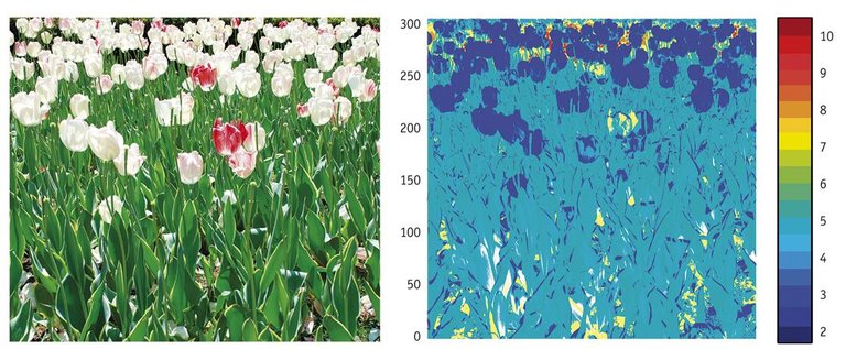
When we work with colors, we believe they can be easily evaluated. We do it at all times: we approach the red notebook or look at the blue car. But for an artificial system this work is very difficult. The first difficulty is that the cameras work only with three colors: red, green and blue; it is called RGB (taking into account the network, green and blue words). And the second is that color is not the unchanging feature of an object. If we see a sheet of paper in the sun it will look white, but if we look in a room with a red light it will look red. When seeing the color the light influences a lot. It is also important what they have around them: in the image of the next page we have two pictures of the same color surrounded by different colors, our eyes tell us that the colors of the picture are different.
With the aim of determining color behavior, researchers have been working for a long time. Newton took his first steps, analyzing how the light decayed through a prism. He is followed by J.C. Maxwell and T. For example, Young followed the same line, to which we owe what we know today about color, which is basically a human perception. That is, color exists because we see the world and associate colors to what we see. The same happens with languages: although a word has its own sound, it makes sense because we give it meaning.
Neuroscience Neuroscience
Being color a characteristic of human perception, we begin to analyze how we see it. For this purpose it was necessary to know the way of working of our visual system. Neuroscience is a discipline that studies our nervous system and in which vision is found. Neuroresearchers start from different approaches: biological, medical, pharmacological... Computational neuroscience studies the way we work in our neurological system to create artificial systems based on biological systems.
Human vision Human vision
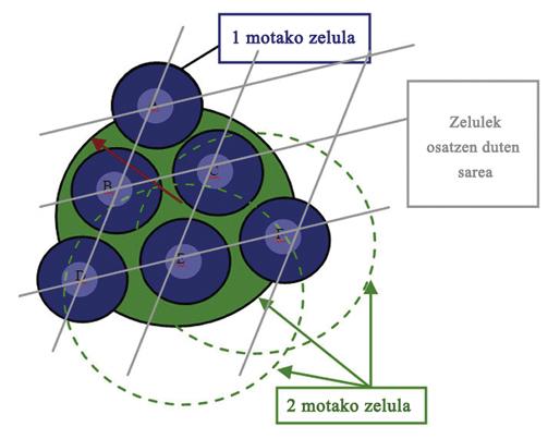
The eyes are the access to human vision. They are a treasure, a treasure that we all have, and just start analyzing it we realize the complexity of the system. Humans have two eyes with more than 100 million photoreceptors (cones and batons). In other words, we have a 100 megapixel camera in each eye, with current digital cameras between 5 and 20 megapixels. But we also have a small computer: the retina. This computer is formed by 6 layers ordered. Each layer of the retina is made by different cells.
Model model Model
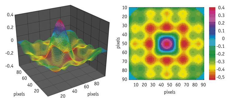
In order to analyze how humans work color, we have developed a mathematical model representing the functioning of the retina. For the construction of this structure we have compiled and analyzed the data of anatomical and neurophysiological research. It begins with the definition of each of the layers that make up the model. We will define the location of each cell of each layer to define the level of cover and the capture zones. In addition, cells are connected to other types of cells, receiving input signals and emitting output signals, simulating dendritic and axonic connections. Finally, the integration of the signals received by the cells and the generation of exit signals by shunts or unifying connections have been other areas that we have worked.
Therefore, in order to build the model, it is necessary to determine where each cell is, what and how it communicates and what type of work it does.
In total, 4 types of cells and 24 subtypes have been taken as a model. The output channels of the system are nodal cells that, once the nerve pulses are encoded, transmit the information through the optic nerve.
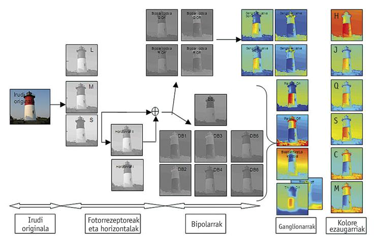
We have defined a system that calculates the color of an image by combining the information channels generated by each group of nodal cells. For this we have based on the calculation of the color attributes proposed by the ciecam02 (Comission Internationale de l'Eclairage 2002). By joining the invented system and the ciecam02a, we have achieved a bioinspired system capable of distinguishing colors the way people distinguish. The color model of an image calculates, for each point, the tone, the whiteness, the luminosity, the saturation, the chroma and the chromatic, as well as the parameters of detection of edges, both in chromatic and non-chromatic components. The mathematical model invented in research work obtained better results than the last model proposed by the CIE when using the Munsell color databases. In addition, the CIE model is used only with laboratory samples, while the model presented is capable of working with real images. The image above shows the images generated by the different cell types of the model, as well as the color characteristics of each pixel. This system opens the doors to advances in artificial vision and improves the results obtained so far. It can be used both to improve the quality of life of people and in industry.
Acknowledgments: Thanks to FUNDACIÓN TECNALIA for its collaboration and thanks to the BASQUE GOVERNMENT for the grant of grants through the ETORTEK program for research at the Massachusetts Institute of Technology and the University of Cambridge.
Bibliography Bibliography Bibliography
Buletina
Bidali zure helbide elektronikoa eta jaso asteroko buletina zure sarrera-ontzian


