


After 3000 years without rest
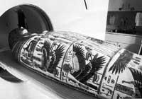
Thanks to CT or computed tomography, well known in medicine, it has been possible to study in detail the body of the woman who died in Thebes 3,000 years ago. This is not the first time a body from the same period has been analyzed, but for the first time the artistic value of the sarcophagus has not been damaged. The use of tomography means that the outer work of the sarcophagus should not be touched or touched and the body hidden by the cover is reformed on a screen.
Researchers at the Toronto Hospital have digitized X-ray images obtained using a tomograph in the scanner and integrated them into the three-dimensional computer program. In this way, the body of the sarcophan woman can be “seen” and “analyzed” without manipulating the exterior of great artistic value. The use of radiation can be disproportionate, as no other damage can occur when dying. This has allowed obtaining images of great precision, which has allowed differentiating sections of different density.
With the use of the tomograph, among other things, you will get a lot of information about the medical techniques used in ancient Egypt. So far it has been clarified that the woman studied in the study died affected by an infection of the orders.
Buletina
Bidali zure helbide elektronikoa eta jaso asteroko buletina zure sarrera-ontzian


