
Whooping cough: a close look
2023/09/01 Amuategi Aulestiarte, Jone - Biokimika eta Biologia Molekularra saila (EHU). Biofisika Institutua (UPV/EHU-CSIC) | Alonso Estrada, Rocio - Biofisika Institutua (UPV-EHU-CSIC), Biofisika Bizkaia Fundazioa | Ostolaza Etxabe, Helena - Biokimika eta Biologia Molekularra saila (EHU). Biofisika Institutua (UPV/EHU-CSIC) Iturria: Elhuyar aldizkaria
Pertussis is a respiratory infection caused by the bacterium Bordetella pertussis and potentially fatal to some populations. Although global immunization campaigns have helped to alleviate the situation, increases occur every 3-5 years. To go further, Osakidetza announced a hearing in Gipuzkoa in June 2023. In this review we have worked on the disease, the bacteria and their virulence factors.
In 2019, lower respiratory tract infections killed 2.4 million people worldwide [38], with children under 5 and older people being the most affected [43]. Those responsible for these infections are viruses and/or pathogenic bacteria, such as the pneumonia bacteria that causes almost half of the deaths [29, 43]. We cannot fail to mention the viruses of the family Coronaviridae, causing the common cold and the diseases SARS, MERS or COVID-19. In this review, in any case, we have seen a whooping cough caused by Bordetella pertussis.
Return of an old disease
Pertussis is a disease with a curious name. However, it disconnects the sound that infected people produce when breathing between a cough episode and the next. Although it may initially look like a conventional cough, symptoms can last for weeks or months. It can cause shortness of breath, sudden episodes of cough and fever, among others. Apnea is also common in infants. If the disease is not overcome in the early stages, in more severe cases it can lead to death from pneumonia in 90% of patients [23].
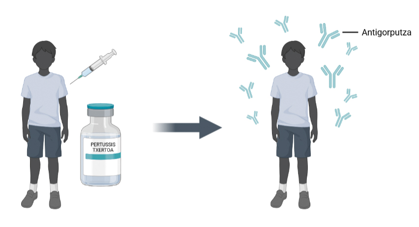
This infection continues to affect and kill many children worldwide, but it can prevent vaccines [16]. According to the latest report published by the World Health Organization (WHO), before pertussis vaccines became available, it was one of the most common childhood diseases. In the 1950s and 1960s, high-level vaccination campaigns led to a 90% reduction in incidence and mortality, at least in industrialized countries (WHO, 2021).
Each country can have its own immunization campaign. For example, the website of the European Centre for Disease Control and Prevention (ECDC) collects information on European programmes. However, at this time, the most common thing is that the triple diphtheria, tetanus and pertussis vaccine is received at once. It is collected during childhood in three doses. According to different global assessments, in 2021 the immune coverage of this vaccine was 81%, the lowest since 2008 [35].
As we have said, vaccines received in childhood are generally very mild. In addition, vaccines for mothers during pregnancy have helped to prevent hospitalization and death of children. In this case, the mother will pass on the antibodies they have created through the placenta, giving protection [23].
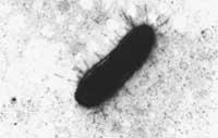
Whooping cough is caused by the bacteria Bordetella pertussis and can be spread by contact with respiratory secretions or saliva droplets. Jules Bordet and Octave Gengou were first identified, isolated and seeded in 1906 [4]. Due to this and other findings related to immunity, Bordet received the Nobel Prize in Medicine and Physiology in 1919.
Although the disease has been known for years, we cannot now overcome it, as the number of people infected increases every 3-5 years. It is currently a problem of public health services in different countries. Thus, for example, the whole of Europe has been in a position since 2011 (ECDC), while in Osakidetza announced a hearing in June 2023 at the Advisory Council.
The truth is that for years the WHO has expressed its concern and recognized that the increase in the disease is occurring worldwide. Unlike other respiratory infections, in this case outbreaks are not uniform, that is, they are not associated with specific sites or stations. Epidemiological studies indicate that the increase may be associated with loss of immunity [22, 20, 34]. In other words, immunity to cucumbers is provisional and therefore expires. It is estimated that natural immunity lasts up to 3.5-30 years, and that acquired by vaccines lasts from 4 to 14 years. Therefore, once the disease has passed, it is very common to re-infect it. However, the severity of symptoms is associated with the time since immunity was acquired [46].
Via cell membranes
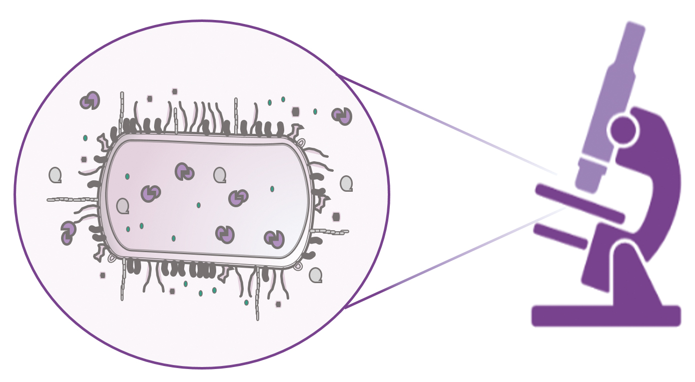
B. pertussis has a specific affinity with the mucosa of the human airways. This means it has the ability to stick on it. To invade and live the upper and lower airways it uses several virulence factors. Some help you adhere to host cells, while others use them to roll the epithelial layer and avoid the immune system. If you want more information about these virulence factors, please read the relevant reviews [15, 9].
The list of folds that this bacterium uses to attack is long. These include porous toxin Adenylate Cyclase, a protein that attacks immune system cells. Thus, it weakens human defenses and the bacteria can spread easily in the body [25, 7].
Pore-forming toxins can be described as biological perforators because they form aqueous channels that adorn the membrane from one side to the other [8, 30]. Biological membranes are a solid and dynamic scaffold to maintain cellular integrity and architecture. They are composed of lipids, proteins and sugars, which separate the internal and external medium from cells [45]. They also regulate what can come in or out of the cell. Therefore, its deterioration may lead to cell death [3, 24, 47]. In all vital realms we can find some organism that produces porous toxin: pathogenic bacteria, nematodes, fungi, parasitic protozoa, frogs, plants and more developed organisms [19]. However, the molecular mechanisms used by all these toxins to attack cells are similar.
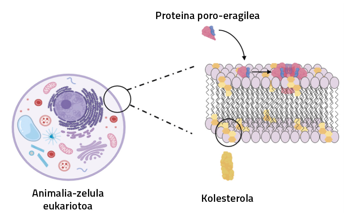
Adenylate cyclase toxin, like other pore-producing proteins, has a special application. At first, when the bacterium produces it and flows to the external environment, it is soluble in water and hides the hydrophobic regions. On the contrary, when the cell meets the host, it changes its appearance: it shows these water-protected regions and is inserted into the membrane [8, 17, 32, 42]. Several proteins are then attached to form the pore, forming a more complex structure. This is how colicins, cytolysins, hemolysins, diphtheria toxin, the protective antigen of anthrax toxin or poro-motor toxins of the Repeats in ToXins (RTX) family work [28]. Pore formation itself is a very dynamic process and therefore difficult to observe. This is a challenge for researchers.
Lipids and proteins, what a pair!
Porotoras toxins, for proper insertion into the cell membrane, are usually accompanied by membrane lipids. Lipids can directly or indirectly adjust the process. On the one hand, they can interact specifically with proteins [26, 36, 4o] and on the other hand, the biophysical characteristics of the membrane (fluidity, phase separation, thickness, tension, etc.). can be modular [10, 21, 39]. Therefore, they can modify the structure and function of the protein.
Membrane cholesterol, for example, controls the activity of multiple membrane receptors through specific interactions. They may also contain proteins such as ATP-Binding Casse receptor receptors and ABC transporters that are specifically aware of cholesterol. These regions are called CRAC or reverse CARC, and although they have very diverse structures, they use similar mechanisms to associate cholesterol. As can be seen in the literature, some amino acids frequently appear in these segments following an already known and described pattern [14, 13].
Many researchers have focused on membrane lipid and protein interactions. It is a topic of great interest that combines scientific disciplines such as biochemistry, biophysics, cell biology or bioinformatics. In recent years, our group has analyzed the interaction between Adenylate Cyclase to xina and membrane, mainly combining biophysics and biochemistry. We have recently demonstrated that the toxic activity of protein depends on membrane cholesterol [18] and that it is a specific interaction between protein and cholesterol [1].
Why basic research?
Humans have learned to live with pertussis and the situation has improved dramatically since vaccines are available to everyone. This is undoubtedly due to basic research and the research plan. The identification, description and understanding of the instruments the bacteria uses to harm humans has been fundamental to explain the molecular mechanisms of the disease and to be able to act against the dome.
Porous toxins have a variable nature depending on the environment. They change very quickly, so it's very difficult to see such dynamic processes. However, they are not isolated mechanisms, but are also produced in the nature in proteins of the vertebrate immune system or in amyloid proteins [27, 37]. Therefore, the effort made to understand these proteins can help other areas.
We have already commented that porous toxins can be really harmful to human health. On the contrary, they have been researched for a long time and can now be useful for future biotechnological applications and so on: targeting cancer cells [31], killing pathogenic bacteria [5], biosensors for detection [2]… It has also been shown that the Adenylate Cyclase toxin can be used as a carrier vaccine [6]. The development of all these applications has first required an understanding of the physical-chemical norms behind these processes, which requires basic research. In addition, collaboration and communication between multidisciplinary researchers will undoubtedly facilitate and expedite the way.
Bibliography
- Amuategi, J., Alonso, R., and Ostolaza, H. 2022. “Four Cholester-Recognition Motifs in the Pore-Forming and Translocation Domains of Adenylate Cyclase Toxin Are Essential for Invasion of Eukaryotic Cells and Lysis of Erythrocytes”. International Journal of Molecular Sciences, 23(15), Article 15. https://doi.org/10.3390/ijms23158703
- Anderluh, G., and Lakey, J. H. 2008. “Disparate use similar architectures to damage membranes proteins”. Trends in Biochemical Sciences, 33(10), 482-490. https://doi.org/10.1016/j.tibs.2008.07.004
- Bernardes, N., and M. Fialho, A. 2018. “Perturbing the Dynamics and Organization of Cell Membrane Components: A New Paradigm for Cancer-Target Therapies”. International Journal of Molecular Sciences, 19(12), 3871. https://doi.org/10.3390/ijms19123871
- Bordet J, Gégou O. 1906. “Pertussis microbe. Ann Inst Pasteur (Paris). 1906;20:731–41.
- Brogden, K. A. 2005. “Antimicrobial peptides: Pore formers or metabolic inhibitors in bacteria?” Nature Reviews Microbiology, 3(3), 238–250. https://doi.org/10.1038/nrmicro1098
- Carneiro, G. B. Castro, J. T., Davi, M., Miyaji, E. N., Ladant, D., and Oliveira, M. L. S. 2023. “Immune responses and protection against Streptococcus pneumoniae elicited by recombinant Bordetella pertussis adenylate cyclase (CyaA) carrying fragments of pneumococcal surface protein A, PspA”. Vaccine. https://doi.org/10.1016/j.vaccine.2023.05.031
- Chenal, A. 2018. Introduction to the Toxins Special Issue on the Adenylate Cyclase Toxin. Toxins, 10, 386.
- Da Peraro, M., and van der Goot, F. G. 2016. Pore-forming toxins: Ancient, but never really out of fashion. Nature Reviews Microbiology, 14(2), Article 2. https://doi.org/10.1038/nrmicro.2015.3
- Dorji, D., Mooi, F., Yantorno, O., Deora, R., Graham, R. M., and Mukkur, T. C. (2018). Bordetella Pertussis virulence factors in the continuing evolution of whooping cough vaccines for improved performance. Medical Microbiology and Immunology, 207(1), 3-26. https://doi.org/10.1007/s00430-017-0524-z
- Escribá, P. V. González-Ros, J. M. Goñi, F. M. Kinnunen, P. C. J. Vigh, L., Sánchez-Magraner, L. Fernández, A. M. Busquets, X. Horváth, I., and Barceló-Coblijn, G. (2008). Membranes: A meeting point for lipids, proteins and therapies. Journal of Cellular and Molecular Medicine, 12(3), 829-875. https://doi.org/10.1111/j.1582-4934.2008.00281.x
- Etymology: Bordetella pertussis. (2010). Emerging Infectious Diseases, 16(8), 1278. https://doi.org/10.3201/eid1608.ET1608
- European Center for Disease Prevention and Control: Pertussis (whooping cough) https://www.ecdc.europa.eu/en/pertussis-whooping-cough 2023-06-19
- Fantini, J., and Barrantes, F. J. (2013). How cholesters interacts with membrane proteins: An exploration of cholvol-binding sites including CRAC, CARC, and tilted domains. Frontiers in Physiology, 4, 31. https://doi.org/10.3389/fphys.2013.00031
- Fantini, J., Di Scala, C., Evans, L. S. Williamson, P. T. F. and Barrantes, F. J. (2016). A mirror code for protein-cholester-interactions in the two leaflets of biological membranes. Scientific Reports, 6(1), Art. 1 https://doi.org/10.1038/srep21907
- Fedele, G., Bianco, M., and Ausiello, C. M. (2013). The virulence factors of Bordetella pertussis: Talented modulators of immune response host. Archivum Immunologiae Et Therapiae Experimentalis, 61(6), 445-457. https://doi.org/10.1007/s00005-013-0242-1
- Fry, N. C., Campbell, H., and Amirthalingam, G. (2021). JMM Profile: Bordetella pertussis and whooping cough (pertussis): still a significant cause of morbidity and mortality, but vaccine-preventable. Journal of Medical Microbiology, 70(10), 001442. https://doi.org/10.1099/jmm.0.001442
- Gilbert, R. J. C. Dalla Serra, M., Froelich, C. J. Wallace, M. I, and Anderluh, G. (2014). Membrane pore formation at protein-lipid interfaces. Trends in Biochemical Sciences, 39(11), 510-516 https://doi.org/10.1016/j.tibs.2014.09.002
- González Bullón, D. Uribe, C. B. Amuategi, J., Martin, C., and Ostolaza, H. (2021). Cholesters stimulates the lytic activity of Adenylate Cyclase Toxin on lipid membranes by promoting toxin oligomerization and formation of pores with a greater effective size. The FEBS Journal, 288(23), 6795-6814. https://doi.org/10.1111/febs.16107
- Gupta, L. C., Molla, J., and Prabhu, A. A. (2023). Story of Pore-Forming Proteins from Deadly Disease-Causing Agents to Modern Applications with Evolutionary Significance. Molecular Biotechnology. https://doi.org/10.1007/s12033-023-00776-1
- Gustafsson L, Hessel L, Storsaeter J, Olin P. Long-term follow-up of Swedish children vaccinated with acellular pertussis vaccines at 3, 5 and 12 months of age indicates the need for a booster do 5 to 7 years of age. Pediatrics. 2006 Sep;118(3):134.ajuste: 10.1542/peds.2005-2746. PMID: 16950988.
- Janmey, P. A., and in Kinnun, P. C. J. (2006). Biophysical properties of lipids and dynamic membranes. Trends in Cell Biology, 16(10), 538-546. https://doi.org/10.1016/j.tcb.2006.08.009
- Jenkinson D. Duration of effectiveness of pertussis vaccine: evidence from 10 year community study. BMJ 1988; 296:612–614.
- Kilgore, P. R. Salim, A. M. Zervos, M. J., and Schmitt, H.-J. (2016). Perussis: Microbiology, Disease, Treatment, and Prevention. Clinical Microbiology Reviews, 29(3), 449-486. https://doi.org/10.1128/CMR.00083-15
- Kulma, M., and Anderluh, G. (2021). Beyond pore formation: Reorganization of the plasma membrane induced by pore-forming proteins. Cellular and Molecular Life Sciences, 78(17), 6229-6249. https://doi.org/10.1007/s00018-021-03914-7
- Ladant, D., and Ullmann, A. (1999). Bordetella pertussis adenylate cyclass: With multiple talent. Trends in Microbiology, 7(4), 172-176. https://doi.org/10.1016/s0966-842x(99)01468-7
- Laganowsky, A., Reading, E., Allison, T. M. Ulmschneider, M. B. Degiacomi, M. T., Baldwin, A. J. and Robinson, C. V. (2014). Membrane bind lipids selectively to modulate their structure and function. Nature, 510(7503), 172-175 https://doi.org/10.1038/nature13419
- Lashuel, H. A., and Lansbury, P. T. (2006). Even amyloid caused by protein aggregates that mimic bacterial pore-forming toxins? Quarterly Reviews of Biophysics, 39(2), 167-201. https://doi.org/10.1017/S0033583506004422
- Lata, C., Singh, M., Chatterjee, S., and Chattopadhyay, K. (2022). Membrane Dynamics and Remodelling in Response to the Action of the Trailers-Damaging Pore-Forming Toxins. The Journal of Membrane Biology, 255(2-3), 161-173. https://doi.org/10.1007/s00232-022-00227-z
- Lukšiic, I., Kearns, P. C., Scott, F., Rudan, I. Campbell, H., and Nair, H. (2013). Viral etiology of hospitalized acute lower respiratory infections in children under 5 years of age—A systematic review and meta-analysis. Croatian Medical Journal, 54(2), 122-134. https://doi.org/10.3325/cmj.2013.54.122
- >Mondal, A. K., and Chattopadhyay, K. (2020). Take Toll on Membranes: Curious Cases of Bacterial β-Barrel Pore-Forming Toxins. Biochemistry, 59(2), 163-170. https://doi.org/10.1021/acs.biochem.9b00783
- Pahle J, Aumann J, Kobelt D, Walther W (2015) Oncoleaking: use of the pore-forming clostridium perfringens enterotoxin (CPE) for suicide gene therapy. Methods Mol Biol 1317:69–85. price: 10.1007/10001-4939-27-2_5
- Parker, M. W., and Feil, S. C. (2005). Pore-forming protein toxins: From structure to function.Progress in Biophysics and Molecular Biology, 88(1), 91-142. https://doi.org/10.1016/j.pbiomolbio.2004.01.009
- Pertussis, WHO: https://www.who.int/health-topics/pertussis#tab=tab_1 (19-06-2023)
- Quinn HE et al. Pertussis epidemiology in Australia over the decade 1995-2005 –trends by region and age group. Communicable Diseases Intelligence, 2007, 31:205–215.
- Rachlin, A., Danovaro-Holliday, M. C. Murphy, P., Sodha, S. V, and Wallace, A. S. (2022). Routine Vaccination Coverage—Worldwide, 2021.
- Renard, K., and Byrne, Morbidity and Mortality Weekly Report, 71(44), 1396-1400. https://doi.org/10.15585/mmwr.mm7144a2B. (2021). Insights into the Role of Membrane Lipids in the Structure, Function and Regulation of Integral Membrane Proteins. International Journal of Molecular Sciences, 22(16), Art. 16 https://doi.org/10.3390/ijms22169026
- Rosado, C. J. et al. (2007). A common fold mediates defense and bacterial attack. Science, 317(5844), 1548-1551. https://doi.org/10.1126/science.1144706
- Safiri, S., Mahmoodpoor, A., Kolahi, A. Nejadghaderi, S. A. Sullman, M. J. M. Mansournia, M. A. Ansarin, K., Collins, G. S. Kaufman, J. S., and Abdollahi, M. (2023). Global burden of lower respiratory infections during the last three decades.Frontiers in Public Health, 10, 1028525. https://doi.org/10.3389/fpubh.2022.1028525
- Room - Estrada, L. A. Leioa, N., Romo, T. D., and Grossfield, A. (2018). Lipids Alter Rhodopsin Function via Ligand-like and Trailers-like Interactions.Biophysical Journal, 114(2), 355-367. https://doi.org/10.1016/j.bpj.2017.11.021
- Sych, T., Levental, K. R., and Sezgin, E. (2022). Lipid-Protein Interactions in Plasma Membrane Organization and Function. Annual Review of Biophysics, 51, 135-156. https://doi.org/10.1146/annurev-biophys-090721-072718
- Surveillance Atlas of Infectious Diseases (ECDC): https://atlas.ecdc.europa.eu/public/index.aspx?Dataset=27etaHealthTopic=38 (2023-06-20)
- Tilley, S. J. and Saibil, H. R. (2006). The mechanism of pore formation by bacterial toxins. Current Opinion in Structural Biology, 16(2), 230-236. https://doi.org/10.1016/j.sbi.2006.03.008
- Troeger et al. Estimates of the global, regional, and national morbidity, mortality, and aetiologies of lower respiratory tract infections in 195 countries: A systematic analysis for the Global Burden of Disease Study 2015. (2017). The Lancet. Infectious Diseases, 17(11), 1133-1161 https://doi.org/10.1016/S1473-3099(17)30396-1
- Vaccine Scheduler (ECDC). Pertussis recommended vaccinations: https://vaccine-schedule.ecdc.europa.eu/Scheduler/ByDisease?SelectedDiseaseId=3etaSelectedCountryIdByDisease=-1 (2023-06-20)
- Watson H. “Biological membranes”. 2015Essays Biochem. 2015;59:43-69. Aj. :10.1042/bse0590043. PMID: 26504250; PMCID: PMC4626904.
- Wearing, H.J., and Rohani, P. 2009. “Estimating the duration of pertussis immunity using epidemiological signatures”. PLoS Patastilla. 5(10), e1000647.
- Zhang, Y. Chen, X., Gueydan, C., and Han, J. 2018. “Plasma membrane changes during programmed cell deaths”. Cell Research, 28(1), Art. 1 https://doi.org/10.1038/cr.2017.133.

Gai honi buruzko eduki gehiago
Elhuyarrek garatutako teknologia