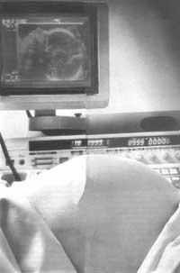
Effects of frequent ultrasound during pregnancy
1994/03/01 Furundarena Salsamendi, Jose Ramon Iturria: Elhuyar aldizkaria
In clinical practice, ultrasounds (used to perform ultrasound) are fully accepted and it has been shown that, if all women are performed between weeks 16 and 20 of pregnancy, perinatal mortality is reduced, mainly due to their ability to detect the malformation of the fetus or the fetus. In addition, they have seen that it serves to determine the age of the fetus, detect growth problems within the uterus, etc.
Doppler technology that uses ultrasound also provides information about blood circulation in the placenta and can serve together with other clinical and biometric data to know why a fetus is small, if it is due to genetic factors or intrauterine pathology.

In a hospital in western Australia, research has been conducted on the effects of frequent use of ultrasound during pregnancy. With 2,834 women, information could be collected since 2,801. They were divided into two groups:
- Group A: The week 18 of pregnancy was ultrasound. In this group, 1,419 women participated.
- Group B: They were ultrasound and Doppler during weeks 18, 24, 28, 34 and 38 of pregnancy. This group was formed by 1,415 women.
Both groups were comparable in terms of age, weight, race, previous pregnancies, tobacco, etc.
As for the results, it has been confirmed that the number of days of hospitalization after birth was the same in both groups, that the deliveries prior to the arrival of the fetus were similar in number and that in many other factors analyzed there were no differences: In the apgar classification, when using intensive responsibilities, when using oxygen, at the age of pregnancy, congenital alterations, etc. In group A there were 10 neonatal deaths and in group B 3.
To know if the weight of newborns is normal, it is compared to a percentile. In this study, below the 10 and 3 percentiles more children appeared in group B and the difference had a statistical significance. However, children in group B weighed only 25 g less than those in group A. The age of pregnancy at the time of birth, the condition of smoker of the parents, obstetric intervention, and the age of the mother also involved a higher risk of appearing below these percentiles.
The doctors who have carried out the research have drawn the following conclusions:
- The frequent use of ultrasound does not improve the results of pregnancy according to the parameters used in this study.
- The occurrence of 10 neonatal deaths in group A (most due to congenital alterations) compared to 3 deaths in group B, is considered random because there are few cases, because neonatal morbidity was similar in both groups and that in those deceased fetuses there were no growth restrictions.
- The fact that in group B more children appear below the low percentiles in weight and then the average weight difference between both groups is so small, would mean that not all fetuses lose weight but that some of them are born with very low percentiles.
- Therefore, they consider that ultrasound should only be used when necessary to complete clinical information during pregnancy.

Gai honi buruzko eduki gehiago
Elhuyarrek garatutako teknologia


