


Intracorporeal images
At present, the blood can be seen moving through the arteries or aspiring a radioactive wall of the muscle heart or locating the blockage in the arteries of a heart that has suffered an attack. You can also see how it moves six months before birth when it is in your mother's stomach. The aim of this article is to review these recently developed techniques. These new techniques are:
CT scan
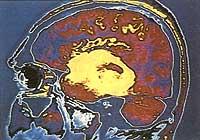
This method was devised in Britain in 1972. This technique basically consists of converting X-ray images into digital computer codes for the realization of high definition video images. The computer graphics used are similar to those used for the realization of distance spatial tests. Showing bone structures in detail, computed tomography highlights small differences between normal and abnormal brain, lung, or other organs.
Despite being a still developing technique, three-dimensional computerized tomography has begun to offer excellent service in reconstructive surgery. Months after the first x-ray photograph by German physicist Wilhelm Konrad Röntgen in 1895, doctors began using X-rays to diagnose bone fractures. In the film appeared dense structures and shadows of the bones that absorbed those mysterious rays. However, soft tissues were easier to cross and were not clear in the images.
Since then, numerous techniques have been developed to obtain cleaner X-ray images. Advanced X-ray machines, by digitizing data, provide accurate images of the tissues.
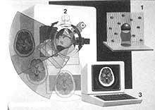
A computerized tomography scanner, crossing the body with a thin X-ray surface, offers a cross-sectional figure of the inner tissues. X-rays that only show the body from an angle can be uninterpretable when the shadows of the bones, muscles, and organs overlap. Large molecules like calcium absorb X-rays when they pass through the body and partially hide what is behind (1).
However, computerized tomography based machines show a section of the body that gives a view taken from multiple angles, rotating a tube that ejects X-rays around the patient (2).
Several sensitive deckers behind the body record what the scanner sees and a computer compares the different views to offer a single video image.
Magnetic resonance imaging
The invention of this revolutionary technique can mean for modern medicine a great advance as that which in 1895 Wilhelm Konrad Röntgen gave in his day with X-rays.
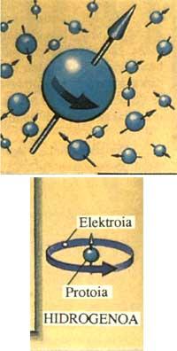
When hydrogen atoms are subjected to the action of a magnetic field, they are aligned. This is the basis of magnetic resonance representation. If a radiofrequency is directed to these atoms, the alignment of their nuclei changes. When the radio waves turn off, the cores re-align and transmit a small electrical signal.
Since the body is formed mainly by hydrogen atoms, it is possible to create an image through pulses that return, showing the tissues and bone marrow as never seen.
Magnetic resonance imaging is expensive. The equipment consists of a large electromagnet, a radiofrequency generator and a computer. The price of this group is 230 million pesetas. In addition, this equipment must be located in a room totally isolated from external radiofrequencies. The cost of the formation of this room can amount to another 112,5 million pesetas.
There are patients who cannot be placed within this magnetic field, such as those who have the posomarator or those who have parts of shrapnel or nails in the body. These metal fragments can be removed from the body by the magnet.
At first they had a lot of concern about the influence that the magnet could have on the human body. They also thought if it could have consequences on human memory. Therefore, at first, in 1974, an onion was inspected. They saw him well the inner rings. In 1977 they first saw a living human fabric: a doll. Two years later, a brave scientist introduced the head into the magnetic field to study his brain.
This technique began to unfold in 1980 and there are currently more than 400 machines working in the US.
For some things, magnetic resonance imaging is more appropriate than computerized tomography. For example, white matter of the brain and gray matter with abundant water. On the contrary, the teeth and bones, having little water, do not appear in magnetic resonance figures. This has its advantages as it allows to see tissues surrounded by bones, such as the spinal cord.
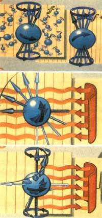
These powerful electromagnets that are used in the manufacture of magnetic resonance imaging are cooled with liquid helium. The high-intensity magnetic field they generate has a great influence on the only proton that forms the nucleus of hydrogen. The axes of the protons that turn in the form of peonzas are usually directed to all directions, without any order. Within the magnetic field of the scanner, however, they are aligned according to the force line. However, although they are aligned, they have a motion of precesion of a certain frequency. The stronger the magnetic field, the higher the frequency.
When the scanner excites the protons with a radio pulse of the same frequency as the motion of precesion, they misalignment describing the circular motion and emitting a radio signal.
A computer converts these signals into images of the scanned area. The figure highlights the density differences of hydrogen atoms. Hydrogen indicates water content, so doctors can use the image to separate the tissues.
Scientists have selected hydrogen as the basis for this technique, as it is a very abundant element in the body and has special magnetic properties. Techniques based on other elements are also being investigated. In fact, the properties of sodium and phosphorus, for example, can report traces of heart attacks.
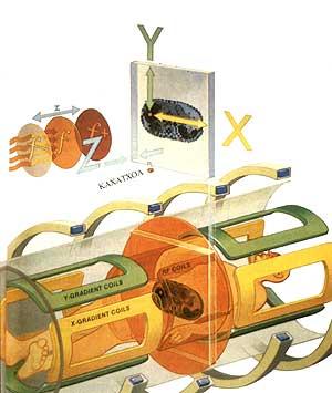
To create the image, the computer creates a three-dimensional network of small boxes. If we call these three dimensions X, Y and Z, first the magnetic field changes direction Z to define a plane from the patient's head to the feet. Within this plane the protons move with a movement of oscillation according to a frequency f. Some coils then pass a radiofrequency pulse. This pulse is of the same frequency as the oscillatory movement of the protons.
Before aligning the protons, other coils modify for a short time the magnetic resistance of the plane in direction Y. This causes protons to describe oscillation movements at different speeds in the downward direction of the plane. Detecting these differences, the computer determines the positions of the boxes in direction Y.
Then the coils modify the magnetic field from left to right in direction X, making the protons correct according to the different frequencies. When the position of each box is determined in the directions X, Y and Z, the computer assigns to each box a point of the screen. The brightness of the point is given by the number of protons inside the box and by the magnetic property of the fabric.
The set of points forms an image. Its ability to show soft tissues with large contrasts makes magnetic resonance imaging an ideal resource for the study of the spinal cord. Doctors who wanted to see the spinal cord before magnetic resonance was accessible had to inject a substance there that contrasted with X-rays. This procedure was dangerous and painful for the patient.
Digital Subtraction Angiography
This technique shows images of blood moving in the blood or of discomfort that make it difficult to progress.
Digital subtraction angiography is based on the injection of a substance in contrast with opaque iodine to X-rays. The shade that generates this opacity allows doctors to see the flow of blood. Often digital subtraction angiography is used to see how the heart is fed with blood. Before injecting the contrast substance, an X-ray image is made that is stored on a computer. After injection, the second image of the blood flow that highlights the substance is made. Then, the computer gets the subtraction of the two images, and a clear picture of the pictures appears.
One of the most frequent surgical procedures at present is the bypass of the cardiac arteries. The blood is offered other alternatives to the arteries that have been blocked by the presence of oily materials or calcifications, by bleeding removed from other parts of the body. In most cases, it is a question of puffs removed from the legs.
With angiography of the digital subtraction and a technique called angioplasty, these interventions can be ruled out.
In coronary artery angioplasty, the doctor inserts a slimmer catheter than pen pain through an artery in the arm or groin. Thanks to the vision offered by digital subst angiography, he directs this catheter into the coronary arteries. Then the contrast substance is injected to obtain the image of the obstruction. Another thinner that enters through the interior of the first catheter carries a balloon to that point. The globit is inflated so that the materials that close the artery and reopen the passage to the blood. An intervention of this type lasts about an hour and a half.
These procedures do not pose danger, are quick, do not produce pain and the recovery of the patient is done quickly. When the plaque that clogs the artery is made of calcified material, the balloon cannot discard it and then runs the risk of interrupting the blood. For patients with this problem at the moment it is best to do bypass.
Digital subtraction angiography is used not only to open barriers that prevent blood from passing, but also to close openings that cause bleeding. To do this, a little gelatin is introduced to stop the bleeding. Also, in these cases, very thin catheters inject some isobutyl–2-cyanoacrylate droplets to close the passage to the blood that goes to the emerging tumors and cut the hemorrhages. This technique has also been used in brain hemorrhages.
Sonography Sonography Sonography
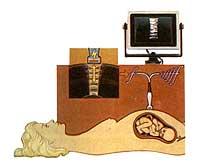
This is a technique created by the development of sonar invented during World War II. For the first time it was used in medicine in the US in the 1950s. A small transducer or transmitter/receiver is placed in contact with the part of the body to be investigated. High-frequency sound waves penetrate the body, collide with the internal organs, and are reflected outward. On return the transducer functions as a receiver. The time taken by the waves in the displacement denounces the position, size, shape and even the texture of the organ and shows them on an online screen.
The most recent advancement of sonography is the digital Doppler color. With the help of the computer, it shows how human blood moves through the heart and blood. Sound or radioelectric waves are reflected in a moving object with a frequency variation. This is the Doppler effect. High-frequency sound waves penetrate the area to investigate, for example in a morcilla, showing the blood flowing into it.
The base of the system is a piezoelectric crystal that transforms electrical pulses into vibrations that penetrate the body. On the return this crystal returns to convert the vibrations that arrive in electrical signals. The doctor places the transducer with crystal on the area to be explored, for example on the belly of a pregnant woman. The echoes reflected by the fetuses become signals that the computer transforms into video images.
Drawing with radioisotope
The two intracorporeal imaging techniques are: the so-called PET (positron emission tomography) and{ (single photon emission computed tomography).
The{ shows blood flow by making images of radioisotope remains. PET, for its part, can measure metabolism by explaining how the body works. The use of tractors with radioisotopes is very appropriate for the investigation of epilepsy, schizophrenia, Parkinson disease and stroke.
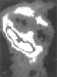
The PET scanner shows how the brain works, showing how local cells consume sugar and other substances.
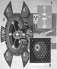
The substance is marked with a radioisotope prepared in a low energy cyclotron. The isotope has a short life, h.d. At the minutes or hours of its creation, it has already lost half of its radioactivity. Once injected into the body, the radioactive solution emits positrons during flow. Positrons collide with electrons, fractions of two types are destroyed each other and two gamma rays are formed, causing a small energy explosion. These two rays come out in opposite directions and touch the crystals of a detector ring that surrounds the patient's head.
The crystals then emit light. A computer determines the position of these light rays and that of the radiation source and converts these data into images. Following the trajectory of the radioactive substance, the doctor can locate areas where abnormal brain activities occur and study cell health.
Like this PET requires the use of a cyclotron, the technique known as{ uses commercial radioisotopes, making it much more economical.
Buletina
Bidali zure helbide elektronikoa eta jaso asteroko buletina zure sarrera-ontzian


