MRI in three dimensions
The objective of the TRAC project is to develop a prototype to visualize the three-dimensional images of the internal organs. To do this, we are working on a technique called augmented reality that, at the end, will serve to plan operations and help in the operating room.
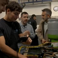
The work is carried out at the Vicomtech research centre. There they work in the field of computer imaging, investigating medical applications, etc. This is where the TRAC project is framed. At the moment, we are working on the increased image of the liver.
From 2D to 3D
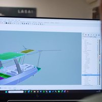
Magnetic resonance scanners give us an image of the body section of 3 to 3 mm. Normally, if it comes to obtaining volume-related information, it is about placing these two-dimensional images together, and the doctor should take advantage of his experience and his ability to interpret the images to form the three-dimensional image of the liver. It is a question of the prototype carrying out this work, generating images in 3 dimensions directly. In the TRAC project, the steps to follow are being worked.
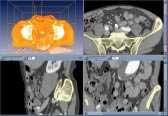
The starting point is the two-dimensional images obtained in conventional magnetic resonance. First of all, these images must be filtered through the appropriate mathematical algorithms by computer. Filters remove electronic noise and highlight details, but you have to choose the right filter to work without losing important nuances.

Then the segmentation algorithms are applied. The aim of these algorithms is to identify the skin of the liver and define the silhouette well. This is one of the most difficult tasks of the project, since the liver is a large, complex organ. Finally, the three-dimensional image is completed with the application of reconstruction algorithms and the obtaining of a volumetric liver image.
This type of images will initially serve to study the external hepatic anatomy and see the extent of the damage, and later, once tightened the system, they will have the sufficient resolution to study the intrahepatic vessels, tumors, etc. But the project has a second part, which joins augmented reality with the operating theatre itself.
Images in the operating room
The objective of this second part is to visualize in the operating room the images obtained by resonance and reorganized in three dimensions, and also placed in the direct image of the patient's body.
You have to take a few steps to get it. On the one hand, a stereoscopic chamber should be placed next to the patient's body. With this special camera, the patient will be filmed in three dimensions during the operation. The images taken by the camera are processed by the computer and are seen on a flat screen with a three-dimensional view.
Likewise, we must know at all times the exact position of the patient's body and stereoscopic chamber. For this purpose, 3 reflective markers are placed on the skin of the patient and another 4 on the stereoscopic chamber. An optical system follows the markers and sends a computer its location. Thus, when moving the patient or stereoscopic camera, the image of the screen also moves.
Finally, three-dimensional images of the liver obtained from magnetic resonance are required. Now, a computer collects all these data and creates an increased view of the patient: the patient's patient and liver are seen simultaneously. And as seen on the special flat screen, they are seen in depth form, as if they had three dimensions. It is intended that these images are also useful in operations.
Vicomtech is responsible for the development and research of the project, but there are more participants: STT, manufacturer and seller of monitoring systems, and BILBOMATICA, specialist in medical databases. Finally, all the results are analyzed by experts of the Cruces Hospital of Bilbao to know their real usefulness. Their doctors are, as it were, in some way, those who give the approval to the product.
Buletina
Bidali zure helbide elektronikoa eta jaso asteroko buletina zure sarrera-ontzian