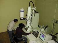
Large Electronic Microscopes
Light is needed to see something in the eye. The light rays that come from any object to the retina have a certain angle, in which the eyes decide that the object may be one size or another. Thus, if with light the light rays reflected in the retina with a large angle, the object would look larger. This happens with the lenses that divert the beam of light that reaches the eyes. The simplest case is that of convex lenses. Optical microscopes are a bit more complex when combining lenses and multiplying thousands of times any object. This way you can see details that cannot be seen visually, such as bacteria. Knowing this, it can be concluded that if you place as many lenses as you want, it would also be possible to reach the smallest detail, but it is not so.
In fact, the radiation wave used to view the object has a limit, you cannot see anything of lesser wavelength than it. It would be like an indivisible rule; if something were measured and greater, is the object equivalent to rules X? could be said, but being the minor object that the rule could not be measured. The same goes for lenses. It cannot be seen when you encounter something that is below the wavelength of the visible electromagnetic spectrum. This wavelength is between the 400-480 nanometers of blue and 620-750 of red. A minor rule is needed to go further. Bands below the wavelength of the auditory band of the electromagnetic spectrum, such as x-rays, can serve this purpose. The wavelength of X-rays is 10,000 times lower than that of visible light, but problems arise because they go through what you want to see; with gamma rays, below the wavelength of the electromagnetic light of vision, the same thing happens.
But there is a highlight and it arises from a principle of quantum physics. According to the principle of wavy corpuscle duality, any object with "p" movement corresponds to the wavelength "L", and as "p" is greater, "L" will be smaller and, in the case of the view, the greater details. Electron microscopes use electrons that not only cross objects but interact with them. The beam of light is usually a beam of electrons and the lenses that can transform their angles are electromagnetic fields, since electrons have electric charge, so electromagnetic fields allow to modify the path of electrons. Viruses can be seen with electronic microscopes, not with optics.
When the electron beam joins the object, three basic types of radiation are created: on the one hand, electrons formed by rebounds after encountering the object, diffuse backwards. On the other hand, electrons coming out of the object after electrons collide with the object are called secondary. And finally, X-rays. Backscatter electrons provide information about the atomic number of the object, X-rays provide information about the chemical structure of the object, and secondary electrons explain the topography of the object's surface, a signal used to visualize the object's image.
How are they?
The most visible part of electronic microscopes is the so-called electron column. Inside the electron column there are:
- Electron cannon with an electron emitting filament. The filament rising releases electrons.
- Electromagnetic lens system. It focuses the electrons released by the filament and takes them to a small diameter. The focusing is done by the electric charge of the electrons, which by their electric charge are exposed to electromagnetic fields and can be transported on a certain path.
- Scanning system. Its goal is to make the targeted electron beam pass over and over again by certain lines on the object surface.
- Detection system. Converts the interaction between the beam and the object into electrical signal.
- Vacuum output. Vacuum is mandatory because if electrons meet an object other than the object, no proper signals will be emitted.
...Electrical signal display systems.
What are they used in?
The scientific basis of the electron microscope, the system it uses to view the images, and once the structure is checked, can also be suitable for what they serve. In fact, the use of electronic microscopes is increasingly widespread: they are used for the realization of veterinary and medical diagnoses, for research in biomedicine, for research in toxicology, for the measurement of particles and by-products, for quality and control processes, for the realization of microstructures of all types of materials with polymers, etc. All said, small things that cannot be seen at first sight, but that will have more and more importance today and in the future.
Published in 7
Buletina
Bidali zure helbide elektronikoa eta jaso asteroko buletina zure sarrera-ontzian