Navarra develops a new tumor limitation tool in medical imaging
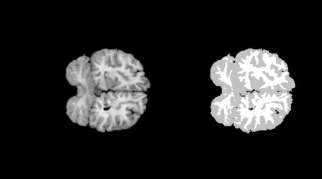
The Artificial Intelligence and Close Reasoning team of the Public University of Navarra (GIARA) has developed a software that automatically limits brain areas in medical images. This work has been awarded by the European Association of Diffuse Logic and Soft Compunting.
"We create machines that simulate man's reasoning, the functioning of the human brain, are software in most cases," explains Humberto Bustince, head of the GIARA group.
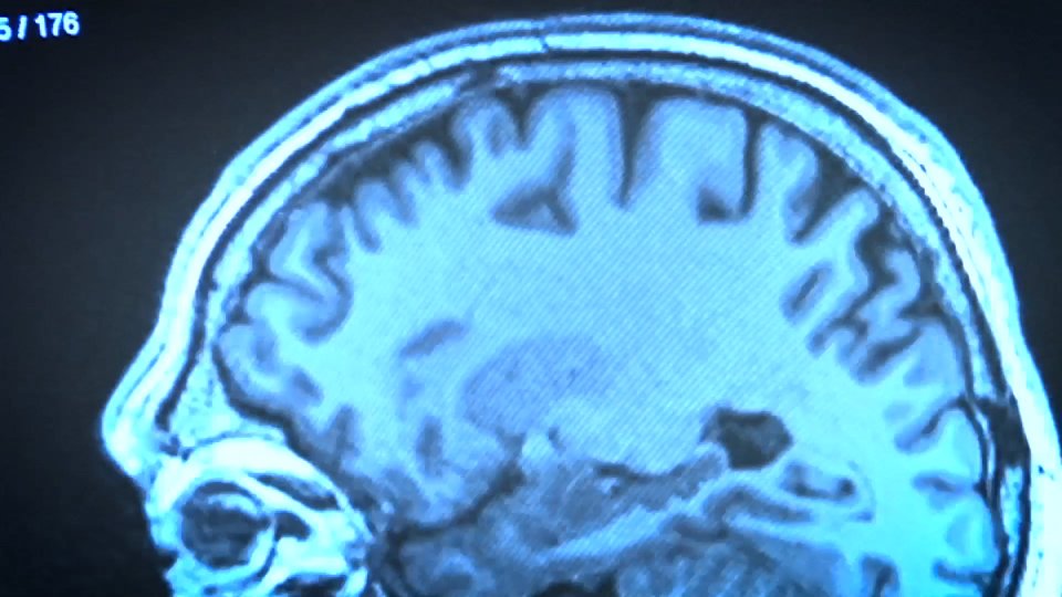
--> See report on the software more specifically Teknopolis helps define tumors in medical images. This allows them to know the state of the diseases. The research focuses on the study of brain images obtained through magnetic resonance.
The program contributes to the process of image segmentation: “The goal is to separate the image from the tumor from the background, behind. At the time of the intervention, it can help the doctor —explains Bustince— to locate exactly the limit of the tumor. Defining this limit is nothing simple and for it we analyze each pixel”.
When completing the image with the data obtained by resonance, the program will select. That is, you must specify the pixels that are part of the tumor. Thanks to mathematics, the software considers the pixels of the image with common features as part of the same object.
“If placed inside the tumor, the last pixel inside the tumor is defined, while if placed outside the tumor, the limit will be the last external pixel of the tumor,” explains Humberto Bustince. These are two different options, and in medicine this situation can be huge. In a brain operation, for example, a difference of three millimeters could mean leaving a person without a voice.”
To the height of the best medical decisions
To measure the effectiveness of the software, the results of the program have been compared with the decisions made by ten physicians on one case. “We can ensure that the option offered by the software is better than the worst decision made by doctors and is up to the best decisions,” added Professor Bustince.
In addition, the algorithm has the advantage of giving the result in real time. In fact, medical images vary in time and often in a short time. “In general, these algorithms are very slow, we have managed to improve the process and the software gives results quickly,” says the research chief of the GIARA group.
The researchers say it is a tool that can help doctors make decisions, but underline that the final decision must always be made by the doctor.
Buletina
Bidali zure helbide elektronikoa eta jaso asteroko buletina zure sarrera-ontzian


